filmov
tv
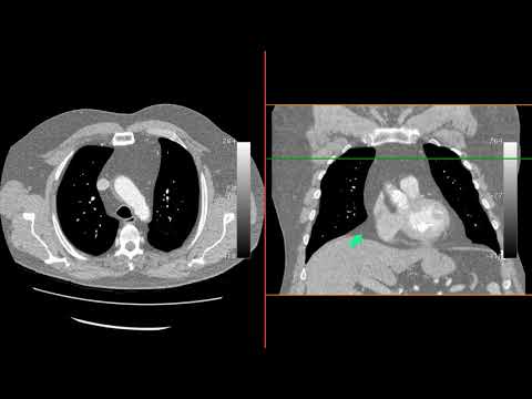
Chest X-Ray breakdown: assessing pericardial calcification

Показать описание
A female in her 60s presents with breathlessness. What does the Chest X-Ray show?
—————————-
CONSTRICTIVE PERICARDITIS
👨🏽💻The first thing to see is that there are bilateral small pleural effusions which in the context of breathlessness raises suspicion of pulmonary oedema
👨🏽💻There is also a ring of calcification outlining the heart raising suspicion of pericardial calcification. Other causes of cardiac calcification we can see on X-Ray include a calcified LV aneurysm, which could be a cause here, as well as aortic valve calcification and mitral annulus calcification
👨🏽💻 In constrictive pericarditis the pericardium becomes thickened and stiff. This causes the pericardium to restrict the normal expansion and contraction of the heart during the cardiac cycle. As a result, there is a disconnect between the pressures inside the chest and those inside the heart
👨🏽💻 During normal respiration, the pressure inside the chest decreases during inhalation, causing blood to flow from the veins into the right atrium and from the lungs into the left atrium. In turn, this increases the volume of blood in the right and left ventricles, allowing the heart to pump more blood to the body
👨🏽💻 However, in constrictive pericarditis, the thickened and stiff pericardium limits the expansion of the heart, preventing it from filling with blood during inhalation. This results in a reduced diastolic filling of the left ventricle. Moreover, the pressure changes inside the chest are not fully transmitted to the left atrium and ventricle, resulting in a dissociation between the intrathoracic and intracardiac pressures
👨🏽💻 This creates a situation where the pressure gradient between the pulmonary capillary wedge pressure and left ventricular diastolic pressure remains constant during the respiratory cycle
👨🏽💻 To compensate for the reduced filling of the left ventricle, the right ventricle fills with more blood during inhalation. The increased blood flow to the right atrium during inhalation is mainly from the inferior vena cava, as it is not affected by the changes in intrathoracic pressure. Additionally, the increased transabdominal pressure during inhalation assists in pushing blood from the inferior vena cava into the right atrium
👨🏽💻 This leads to an increased jugular venous pressure during inhalation, which is known as Kussmaul's sign. During exhalation, the opposite changes occur in the filling of the left and right sides of the heart
@theradiologistpage
✔️ Patient consent obtained
#theradiologist #radiology #radiologist #physician #physicianassistant #medicine #medstudent #medicalstudent #medschool #medicalschool #radtech #mri #medical #radiography #radiologystudent #doctor #medicalstudents #interventionalradiology #cardiacmri #surgeon #medic #frcr #cardiology #nursepractitioner #usmle #medlife #exams #premed
 0:02:41
Chest X-Ray breakdown: assessing pericardial calcification
 0:02:01
Chest X ray in Constrictive Pericarditis (Part 7)
 0:02:14
Chest X-Ray breakdown: how to assess mediastinal contour
 0:02:01
constrictive pericarditis
 0:05:23
Pericardial and cardiac calcification (non coronary artery)
 0:00:16
Pericardial calcification.
 0:09:36
Acquired Cardiac Disease: Unequal Chamber Dilation
 0:12:20
Heart Conditions: Chest X-rays Made Easy
 0:00:21
CXR sign - Tubular artery sign- Pneumomediastinum
 0:02:23
Pseudopleural plaque
 0:01:23
Chest X-Ray breakdown: male presents with cough
 0:00:56
Mediastinal Widening in Chest Pain
 0:12:43
A missed, uncommon lesion on a chest radiograph
 0:04:20
Azygoesophageal Recess #radiology #xray #thoracicsurgery
 0:40:42
Pleural Disorders | Chest Radiology Essentials
 0:13:12
Quick Review of Chest X-Rays
 0:00:28
CXR - Hilar Overlay Sign - Exclusion of middleMediastinal Mass - Radiology Review
 0:02:24
Chest X-Ray breakdown: haemoptysis in a young male
 0:57:30
Session 18: ' Approach to Chest X-Rays in Neonates ' by Dr. Aparna C. | KIMS Cuddles O-N-E
 0:00:56
HILUM OVERLAY SIGN
 0:17:33
How to Interpret a Chest X Ray Lesson 5 Cardiac Silhouette and Mediastinum
 0:01:01
CXR - Encysted pleural fluid
 0:13:30
How to read chest CT: mediastinal anatomy
 0:17:43
Radiology Case Conference
Комментарии