filmov
tv
Roth Spots.

Показать описание
🔷Roth Spots🔷
🟢 White centered retinal hemorrhages, also known as Roth spots, were first described by Moritz Roth, a Swiss physician in 1872.
🟢 However, it was not until 1878 that this condition was assigned the name "Roth spot" by Moritz Litten.
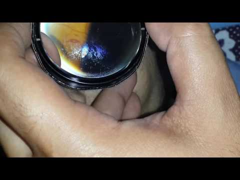
🟢 Roth spots are seen on fundoscopic examination and tend to be located in the peripheral or posterior poles of the eyes.
🟢 Retinal endothelial dysfunction appears to be a common thread among the diverse causes of white centered retinal hemorrhage.
🟢 White centered retinal hemorrhages are a nonspecific ophthalmologic finding seen in multiple systemic conditions of various etiologies.
🟢 Roth spots are most commonly associated with infective endocarditis and have been detected in 80 percent of cases of subacute bacterial endocarditis.
🟢 The differential diagnosis of Roth spots is vast and includes:
👉 Bacterial endocarditis
👉 Anemia/thrombocytopenia
👉 Collagen vascular disease
👉 Leukemia
👉 Hypertensive retinopathy
👉 Diabetic retinopathy
👉 Pre-eclampsia
👉 Human Immunodeficiency Virus (HIV)
👉 Anoxia
👉Shaken baby syndrome
👤 J.R. Fontenla, MD-PhD.
Medical Consultant in Ophthalmology
🏨 Hospital Clinic of Barcelona
Professor of Ophthalmology
🏛 University of Barcelona
I hope you find this video helpful. If you do, please like and subscribe for more videos like this one!
**MEDICAL LEGAL DISCLAIMER**: Visus Formación en Oftalmología does not provide medical advice, and the information available on this channel does not offer a diagnosis or advice regarding treatment. Information presented in these lessons is for educational purposes ONLY, and information presented here is not to be used as an alternative to a healthcare professional’s diagnosis and treatment of any person/animal. Only a physician or other licensed healthcare professional are able to determine the requirement for medical assistance to be given to a patient. Please seek the advice of your physician or other licensed healthcare provider if you have any questions regarding a medical condition.
*IMAGE DISCLAIMER: The content (ex. images) used in this lesson are used in accordance with Fair Use laws and are intended for educational/teaching purposes only*
🟢 Check out some of our other lessons:
Channel Home Page
English Content Playlist
Retina Playlist
Vitreous Playlist
Neuro-Ophthalmology Playlist
Glaucoma Playlist
Cornea Playlist
Conjunctiva Playlist
Uvea Playlist
Lens Playlist
Sclera Playlist
Eyelids Playlist
Orbit Playlist
Lacrimal System Playlist
🟢 Being a Channel Member makes you our Partner. A Partner that contributes to the maintenance of the channel and the creation of new educational/teaching content.
🟢 Being a Member 😎 also gives you exclusive access to:
👉 Videos with additional content.
👉 Multiple Answer Question Bank.
👉 Diagnostic Photo Bank and Photos of clinical cases.
🤩 If you want to become our Partner and contribute to the Content Creation and Maintenance of the Channel, become a member.
Thank you!! 😀👍.
🟢 White centered retinal hemorrhages, also known as Roth spots, were first described by Moritz Roth, a Swiss physician in 1872.
🟢 However, it was not until 1878 that this condition was assigned the name "Roth spot" by Moritz Litten.
🟢 Roth spots are seen on fundoscopic examination and tend to be located in the peripheral or posterior poles of the eyes.
🟢 Retinal endothelial dysfunction appears to be a common thread among the diverse causes of white centered retinal hemorrhage.
🟢 White centered retinal hemorrhages are a nonspecific ophthalmologic finding seen in multiple systemic conditions of various etiologies.
🟢 Roth spots are most commonly associated with infective endocarditis and have been detected in 80 percent of cases of subacute bacterial endocarditis.
🟢 The differential diagnosis of Roth spots is vast and includes:
👉 Bacterial endocarditis
👉 Anemia/thrombocytopenia
👉 Collagen vascular disease
👉 Leukemia
👉 Hypertensive retinopathy
👉 Diabetic retinopathy
👉 Pre-eclampsia
👉 Human Immunodeficiency Virus (HIV)
👉 Anoxia
👉Shaken baby syndrome
👤 J.R. Fontenla, MD-PhD.
Medical Consultant in Ophthalmology
🏨 Hospital Clinic of Barcelona
Professor of Ophthalmology
🏛 University of Barcelona
I hope you find this video helpful. If you do, please like and subscribe for more videos like this one!
**MEDICAL LEGAL DISCLAIMER**: Visus Formación en Oftalmología does not provide medical advice, and the information available on this channel does not offer a diagnosis or advice regarding treatment. Information presented in these lessons is for educational purposes ONLY, and information presented here is not to be used as an alternative to a healthcare professional’s diagnosis and treatment of any person/animal. Only a physician or other licensed healthcare professional are able to determine the requirement for medical assistance to be given to a patient. Please seek the advice of your physician or other licensed healthcare provider if you have any questions regarding a medical condition.
*IMAGE DISCLAIMER: The content (ex. images) used in this lesson are used in accordance with Fair Use laws and are intended for educational/teaching purposes only*
🟢 Check out some of our other lessons:
Channel Home Page
English Content Playlist
Retina Playlist
Vitreous Playlist
Neuro-Ophthalmology Playlist
Glaucoma Playlist
Cornea Playlist
Conjunctiva Playlist
Uvea Playlist
Lens Playlist
Sclera Playlist
Eyelids Playlist
Orbit Playlist
Lacrimal System Playlist
🟢 Being a Channel Member makes you our Partner. A Partner that contributes to the maintenance of the channel and the creation of new educational/teaching content.
🟢 Being a Member 😎 also gives you exclusive access to:
👉 Videos with additional content.
👉 Multiple Answer Question Bank.
👉 Diagnostic Photo Bank and Photos of clinical cases.
🤩 If you want to become our Partner and contribute to the Content Creation and Maintenance of the Channel, become a member.
Thank you!! 😀👍.
 0:03:52
 0:08:45
 0:00:32
 0:00:10
 0:02:50
 0:02:43
 0:40:06
 0:01:01
 0:00:50
 0:02:38
 0:00:58
 0:00:24
 0:01:00
 0:00:47
 0:56:33
 0:00:36
 0:00:11
 0:00:51
 0:00:05
 0:00:40
 0:00:05
 0:02:15
 0:00:12
 0:00:17