filmov
tv
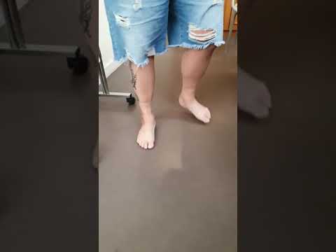
59 Yo male with Charcot Ankle Fracture

Показать описание
A 59 yo male presented to us exactly how you see pictured. He was 6 weeks out from bi-malleolar ORIF — he was instructed by the index surgeon he could bear weight in a walking boot as tolerated due to the 6 week x-rays showing good alignment & what they felt was interval healing. Unfortunately, the neuropathic DM went home, & walked on the right ankle without boot on, converting himself to an open Charcot ankle fracture. The patient as stated, presented to us after the fibula plate had been removed, & had been debrided. …. Timing was tricky as I was leaving town the day after patient presented, so I asked my partner Clay Strong, MD to take the patient to the OR, & try to achieve closure prior to what obviously would be definitively treated with a TTC arthrodesis.
Dr. Strong placed the patient in delta frame you see pictured, excised fibula, closed the lateral wound, & placed a wound vac. The patient was directly admitted from our office to hospital. A PICC line was placed, & he was started on antibiotics (although every culture taken from the lateral plate removal, to the TTC arthrodesis was negative).
-After approximately 15 days in the delta frame, lateral incision looked healthy, labs were normal (aside from A1C of 9) (& small wound at tip of ipsilateral great toe was healed), patient was scheduled for TTC with the Dynanail.
-To avoid compromising the lateral tissues given multiple iatrogenic violations we elected to use medial approach — this also gave us access to remaining HW.
-Due to the amount of vascularity provided by the medial mall & surrounding envelope we elected not to excise it, despite the risks associated with index hardware being placed there. The open fracture was treated early, & aggressively by both us, & the index surgeon & yielded neg cultures.
-Fixation of the medial mall certainly may be criticized. We used a screw designed for the nail which only offered decent compression. We wanted something fully threaded due to cancellous nature of the bone, & recent screw holes.
-The CT was performed at closer to 11 wks than 10, & demonstrates uniting bone. Pt is now 12+ wks from DOS, & transitioning to shoes w/ orthosis for mild varus.
 0:00:22
 0:00:06
 0:00:05
 0:00:53
 0:00:47
 0:00:21
 0:02:11
 0:00:16
 0:01:36
 0:00:08
 0:01:00
 0:01:16
 0:01:07
 0:01:01
 0:00:06
 0:05:55
 0:00:18
 0:00:55
 0:04:16
 0:00:28
 0:03:50
 0:11:11
 0:02:59
 0:22:18