filmov
tv
OCT: Interpreting the image

Показать описание
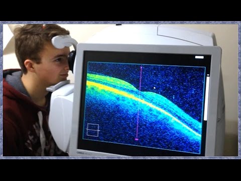
Optical coherence tomography (OCT) is used increasingly in optometric practice to identify retinal pathology, to improve referral accuracy and to monitor for progression of disease. It is vital that the optometrist understands the image in front of them and knows how to describe any abnormality to another professional. Brought to you with Heidelberg Engineering Ltd, this video shows a systematic approach to interpreting the OCT, use of terminology and when to refer. We use case examples and explore what happens in the hospital setting.
Chapters
0:00 CET Learning Objectives
0:44 Introduction
4:55 Normal OCT
6:05 Layers of the retina
9:52 Evaluating OCT images
17:18 What to look for
17:41 Terminology
19:00 Additional Structures
19:23 Systematic procedure
22:28 Final thoughts
23:12 Commonly occurring chorio-retinal pathologies
23:30 Case 1 - AMD
25:50 Case 2 - Diabetic retinopathy
30:40 Anti-VEGF
33:27 Case 3 - Gross macular oedema
35:25 Myopic choroidal neovascularization
36:00 Case 4 - Macular hemorrhage
37:39 Central serous retinopathy
40:56 Vitreomacular interface disorders
41:21 Epiretinal membrane
42:14 Vitreomacular traction
40:02 Case 5 - Macular hole
48:44 Retinal vein occlusion
48:55 Branch retinal vein occlusion
49:19 Central retinal vein occlusion
49:54 Case 6 - Branch retinal vein occlusion
51:10 Case 7 - Macular hemorrhage
52:55 Case 8 - Central retinal vein occlusion + treatment
55.02 Retinal artery occlusion
57:00 Valsalva retinopathy
57:29 Case 9
58:30 Late onset Coats
OCT: Interpreting the image
 0:56:58
Ordering and Interpreting Retinal OCT Images
 0:07:12
OCT Image Interpretation
 0:50:07
Macular OCT Interpretation: A Practical Discussion with Dr. David E. Lederer
 0:13:53
HOW TO READ MACULAR OCT PRINTOUT? made easy!!
 0:30:42
OCT (Optical Coherence Tomography)- Elias Hanna, Univ of Iowa
 0:11:06
OCT-guided PCI: step-by-step_Shlofmitz_Coronary On Demand
 0:16:45
Systematic Interpretation of OCT Angiography Images
 0:04:51
OCT Interpretation - Vitreous
 0:15:56
10 Hacks for OCT Interpretation in Glaucoma - Dr. Mark Dunbar
 0:03:59
Understanding Optic Nerve OCT
 0:31:32
WHAT IS OCT || OCT technology explained ||
 0:00:17
Normal oct vs Abnormal oct retina #shortvideo #viral #knowledgewithani
 0:23:38
HOW TO READ AN OCT PRINTOUT IN GLAUCOMA || BASIC TESTING PROTOCOLS|| ( RNFL, ONH & MACULAR ANALY...
 0:06:19
Using OCT in Glaucoma - Tutorial for Beginners
 0:03:46
What is OCT Scanning? (Optical Coherence Tomography)
 1:30:19
How to Read an OCT Image - with Dr. Jerome Sherman
 0:19:46
OCT Image Interpretation: An Expert Perspective by Dr. Takashi Akasaka
 1:12:44
Interpretation of OCT Retina from A-Z
 0:03:22
OCT line scan interpretation - Inner Retina
 0:19:27
HOW TO READ AN OCT PRINTOUT IN GLAUCOMA || STEPWISE || EXTREMELY EASY || PART -2
 0:00:31
What is an OCT test?
 0:28:28
OPTICAL COHERENCE TOMOGRAPY (OCT) MACULA INTERPRETATION
 0:45:46
Getting Started With OCT - Ocular Coherence Tomography
Комментарии