filmov
tv
Aliasing Artifact | Ultrasound Physics Course | Radiology Physics Course #23

Показать описание
*High yield radiology physics past paper questions with video answers*
Perfect for testing yourself prior to your radiology physics exam 👇
=========================
*I have also created two RADIOPAEDIA LEARNING PATHWAYS*
WHAT’S INCLUDED?
✅This YouTube series Ad free
✅Constantly updated Radiopaedia articles
✅Summary slides
✅Key take home bullet points throughout
✅Multiple review quizzes
✅Short answer review questions
✅Official Radiopaedia course completion certificate
=========================
Aliasing occurs when the pulse repetition frequency is less than twice the doppler shift frequency of the returning echo. This limit is known as the Nyquist limit. In this talk we review how aliasing occurs and what we can do to reduce aliasing in an image. We will review the determinants of pulse repetition frequency and the doppler equation and how these parameters influence the degree of aliasing in a spectral doppler ultrasound waveform.
=========================
*Not sure if the question banks are for you?*
If you're here, you're likely studying for a radiology physics exam. I've spent the last few months collating past papers from multiple different countries selecting the most commonly asked questions. You'll be surprised how often questions repeat themselves!
The types of questions asked in FRCR, RANZCR AIT, ARRT, FC Rad Diag (SA), ABR qualifying Core Physics and MICR part 1 are surprisingly similar and the key concepts remain the same throughout. I've taken the most high-yield questions and answered them in video format so that I can take you through why certain answers are correct and others are not.
Happy studying,
Michael
#radiology #radres #FOAMrad #FOAMed
 0:15:12
Aliasing Artifact | Ultrasound Physics Course | Radiology Physics Course #23
 0:01:20
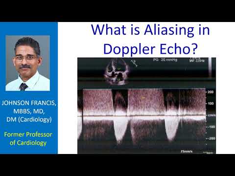
What is Aliasing in Doppler Echo?
 0:00:59
Aliasing vs Doppler Shift
 0:12:45
Color Doppler Artifacts and Spectral Doppler Artifacts
 0:02:49
Aliasing
 0:01:06
Aliasing Ultrasound Physics
 0:04:56
Aliasing and Nyquist Limit in Doppler Echocardiography
 0:00:44
What is Aliasing in Doppler Echo?
 0:06:35
Ultrasound Artifacts of Physics
 0:23:08
Spectral Doppler Ultrasound | Ultrasound Physics Course | Radiology Physics Course #22
 0:17:38
Ultrasound Artifacts | Ultrasound Physics Course | Radiology Physics Course #25
 1:30:20
Unit 20: Doppler Application
 0:38:16
Doppler Ultrasound 101 | The Basics
 0:01:01
Aliasing explained #VeritasiumContest
 0:05:26
Understanding Pulse Repetition Frequency, Scale, Wall Filter and Nyquist Limit.
 0:00:28
Mirror Imaging Doppler Artifacts
 0:03:29
What is aliasing and the Nyquist theorem?
 0:34:15
Aliasing (Wraparound) Artifact and Parallel Imaging in MRI | MRI Physics Course #13
 0:02:49
Color Flow Doppler Direction Tutorial
 0:03:06
Aliasing effect
 0:01:11
Refraction Artifacts
 0:22:32
Doppler Principles
 0:24:47
Continuous vs Pulsed Wave Doppler Ultrasound | Ultrasound Course | Radiology Physics Course #21
 0:00:47
Ghosting Artifact
Комментарии