filmov
tv
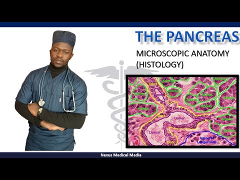
Microscopic anatomy of the pancreas

Показать описание
Under the microscope we can look at the classic histology of the insulin producing islets of Langerhans and see how the exocrine cells of the pancreas are arranged around them to send all of those pancreatic digestive juices to the small intestine.
Music:
Disco 93 by Gregory David
Music:
Disco 93 by Gregory David
 0:22:52
Microscopic anatomy of the pancreas
 0:06:12
Pancreas: Histology
 0:04:40
Pancreas |Histology (Microscopic Anatomy)
 0:07:43
Histology of Pancreas
 0:02:59
Histology of Pancreas
 0:04:13
Histology of the Pancreas: Endocrine and Exocrine
 0:17:31
Pancreas Structure and Function | Digestive System
 0:00:23
Pancreas under the microscope #pancreas
 0:03:15
Histology - Pancreas
 0:01:13
Identification of histology slide : how pancreas looks under microscope
 0:00:16
real nervous system #anatomy #human #body
 0:06:34
Gross anatomy of the pancreas
 0:02:24
The Pancreas under light microscope.
 0:01:13
Microscopic Pancreas
 0:09:08
Pancreas Functional Anatomy
 0:08:54
Pancreas: Mucinous cystic neoplasm - Microscopy (Talking slide)
 0:00:18
REAL Human Pituitary Gland
 0:00:23
How you think the nervous system is😳 #shorts
 0:08:26
Endocrine histology
 0:00:16
Human pancreas (T.s) Under Microscope
 0:39:31
Anatomy of Pancreas (Made Easy)
 0:11:14
Lecture On Microscopic Structure Of Pancreas
 0:07:51
Pancreas and its functions
 0:07:08
Histology with Chimmalgi: Pancreas
Комментарии