filmov
tv
How to Interpret a Chest X-Ray (Lesson 10 - Self Assessment): Part 1
Показать описание
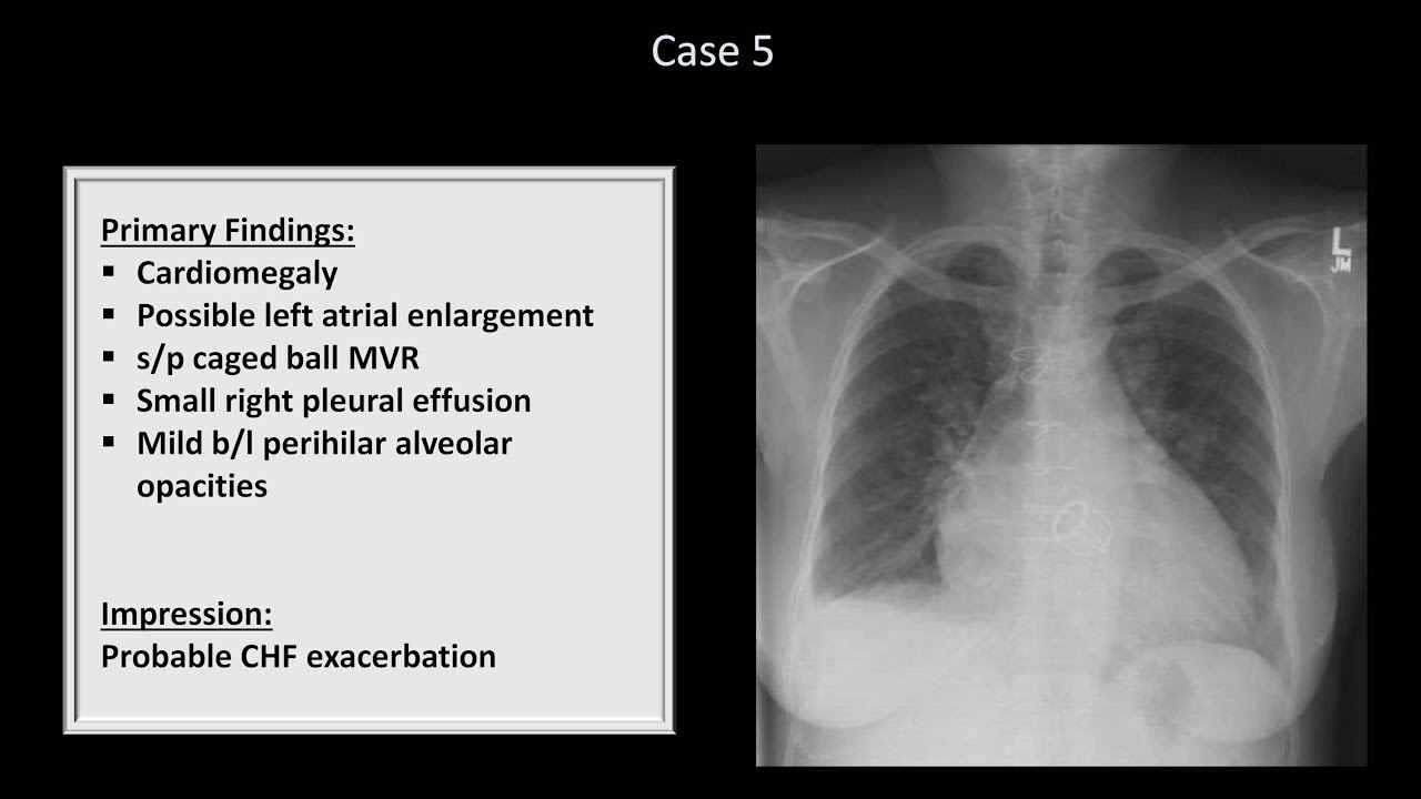
A self-assessment of how much material was learned and retained from this course on interpreting chest x-rays, and how well it can be incorporated into the viewer's preexisting clinical knowledge.
 0:10:11
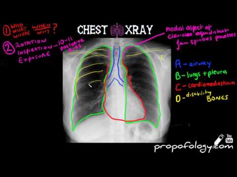
How to Interpret a Chest X-Ray (Lesson 2 - A Systematic Method and Anatomy)
 0:07:02
Reading a chest X-ray
 0:10:30
Chest X Ray Interpretation Explained Clearly - How to read a chest Xray
 0:14:24
How to Interpret a Chest X-Ray (Lesson 1 - An Introduction)
 0:12:37
Chest X Rays (CXR) Made Easy! - Learn in 10 Minutes!
 0:19:44
How to read a chest X-ray (in 20 mins) !
 0:19:19
How to Interpret a Chest X-Ray (Lesson 10 - Self Assessment): Part 1
 0:17:21
How I Read a Chest CT
 0:03:54
How to interpret a Chest Xray in under 4 minutes
 0:05:54
LEARN to Read a Chest Xray in 5 minutes!
 0:16:56
How to Interpret a Chest X-Ray (Lesson 4 - Airways, Bones, and Soft Tissues)
 0:11:42
Chest X-Ray Interpretation Explained Clearly - How to read a CXR
 0:07:19
How to Read a Chest X-ray like a Radiologist! (My Search Pattern)
 0:24:44
Chest X-ray Anatomy | Radiology anatomy part 1 prep | How to interpret a chest X-ray
 0:23:20
How To Read A Chest X-ray For Beginners - Dr. Gill
 0:17:33
How to Interpret a Chest X-Ray (Lesson 5 - Cardiac Silhouette and Mediastinum)
 0:10:30
Chest X ray interpretation (in 10 minutes) for beginners🔥🔥🔥 #chestxray #cxr
 0:16:34
How to Interpret a Chest X-Ray (Lesson 3 - Assessing Technical Quality)
 0:03:46
Anatomy of a Chest X-Ray - How to Read a Chest X-Ray (Part 1)
 0:16:57
How to Interpret a Chest X-Ray (Lesson 7 - Diffuse Lung Processes)
 0:17:51
How to Interpret a Chest X-Ray (Lesson 10 - Self Assessment): Part 2
 0:17:41
How to Interpret a Chest X-Ray (Lesson 6 - Diaphragm and Pleura)
 0:36:54
Introduction to CT Chest - Anatomy and Approach
 0:05:25
How to Read and Interpret Chest X-Rays: OUR TOP 10 TIPS
Комментарии