filmov
tv
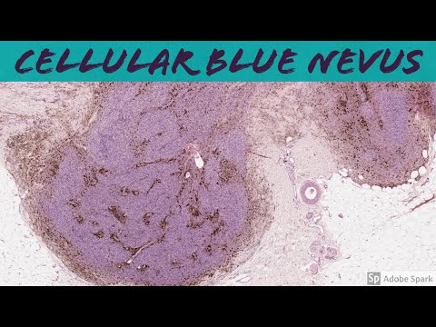
Cellular Blue Nevus: 5-Minute Pathology Pearls

Показать описание
This video is geared towards medical students, pathology or dermatology residents, or practicing pathologists or dermatologists. Of course, this video is for educational purposes only and is not formal medical advice or consultation.
Presented by Jerad M. Gardner, MD. Please subscribe to my channel to be notified of new pathology teaching videos.
Follow me on:
Snapchat: JMGardnerMD
Twitter: @JMGardnerMD
Instagram: @JMGardnerMD
 0:04:10
Cellular Blue Nevus: 5-Minute Pathology Pearls
 0:04:30
Blue Nevus (pathology dermpath dermatology dermatopathology)
 0:39:50
Melanocytic Dermpath: Cellular Blue Nevus vs Deep Penetrating Nevus/Melanocytoma
 0:03:34
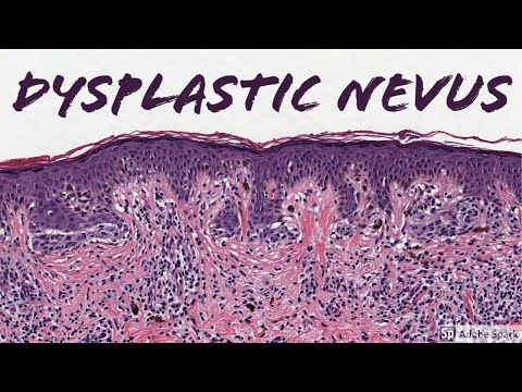
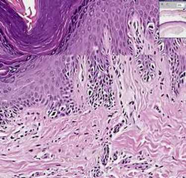
Dysplastic Nevus: 5-Minute Pathology Pearls
 1:10:27
Spitz Nevus, Dysplastic Nevus, Blue Nevus, Congenital Nevus & more - Dermatopathology Basics
 0:05:29
Superficial Spreading Melanoma: 5-Minute Pathology Pearls
 0:04:25
Melanoma vs Nevus: Microscopic Clues for Malignancy Explained in 5 Minutes
 0:05:37
Melanoma (Lentigo Maligna Type) vs Dysplastic Nevus: 5-Minute Pathology Pearls
 0:03:20
Merkel Cell Carcinoma: 5-Minute Pathology Pearls
 0:16:05
Dermpath Made Simple Benign Nevi
 0:04:13
Histopathology Skin Pigmented Spindle Cell Nevus
 0:05:54
Clues to the diagnosis of dendritic and cellular blue nevus (#NAIP)
 0:05:33
Histopathology Skin--Melanoma in situ
 0:02:03
Warty Dyskeratoma: 5-Minute Pathology Pearls
 0:04:57
Desmoplastic Melanoma: 5-Minute Pathology Pearls
 0:03:56
Pathology Highlights : Combined deep penetrating nevus
 0:03:50
Leiomyoma, Pilar Type (Piloleiomyoma): 5-Minute Pathology Pearls
 0:04:19
Atypical Fibroxanthoma (AFX) vs Pleomorphic Dermal Sarcoma (PDS): 5-Minute Pathology Pearls
 0:04:53
Juvenile Xanthogranuloma (JXG): 5-Minute Pathology Pearls for Dermatopathology & Dermatology
 0:01:27
Syringoma: 5-Minute Pathology Pearls (Tadpole / Paisley Tie Pattern Dermpath Dermatology)
 0:05:52
A 6th finger?! Supernumerary (Accessory) Digit: 5-Minute Pathology Pearls
 0:08:12
Pitfalls in the diagnosis of pigmented lesions
 0:03:36
Rosai Dorfman Disease: 5-Minute Pathology Pearls for Dermatology & Dermatopathology
 0:02:32
Mastocytosis: 5-Minute Pathology Pearls
Комментарии