filmov
tv
filariasis

Показать описание
A parasitic disease caused by the superfamily Filarioidea (roundworm).
(Groups)
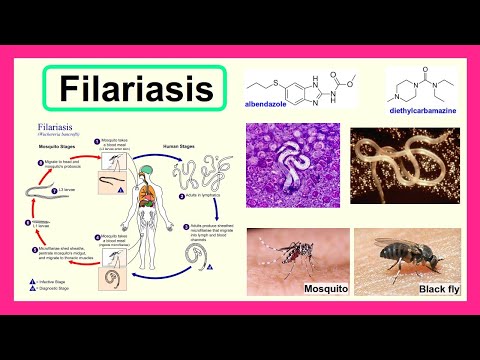
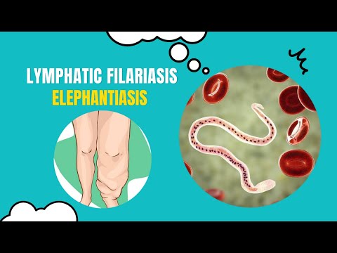
• Lymphatic filariasis: Caused by Wuchereria bancrofti, Brugia malayi, and Brugia timori, leading to elephantiasis. They occupy the lymphatic system.
• Subcutaneous filariasis: Caused by Loa loa (the eye worm) leading to Loa loa filariasis, Mansonella streptocerca, and Onchocerca volvulus leading to river blindness. They occupy the layer just under the skin.
• Serous cavity filariasis: Caused by Mansonella perstans and Mansonella ozzardi. The infections are often asymptomatic, they can be associated with angioedema, recurrent pruritic subcutaneous lesions, fever, headaches, arthralgia, and neurologic manifestations. They occupy the serous cavity of the abdomen.
(Transmission)
The adult worms, which usually stay in one tissue, release early larval forms (microfilariae) into the blood, that can be taken up by blood-feeding insects (e.g. black flies, mosquitoes), where they develop into infective larvae that can be spread to another person.
(Diagnosis)
• concentration methods: Separate the parasites from fecal debris.
• eosinophilia: A nonspecific primary sign.
• finger prick test: Identifies microfilariae on Giemsa stained, thin and thick blood film smears. Blood must be drawn at appropriate times, which reflect the feeding activities of the vector insects. Examples are W. bancrofti, whose vector is a mosquito, night is the preferred time for blood collection. Loa loa's vector is the deer fly, daytime collection is preferred.
• DEC provocation test: The patient is given a single oral dose of diethylcarbamazine (DEC), followed by a blood sample 30-45 minutes later. This can push microfilaria into the peripheral blood during day time which has a sensitivity almost comparable to that of night blood surveys.
• skin snips: Superficial skin biopsies taken from the skin around the iliac crest. M. streptocerca and O. volvulus produce microfilariae that do not use the blood. They reside in the skin only.
• polymerase chain reaction (PCR): Detects filarial antigens in the blood.
• antigenic assays: Detects filarial antigens in the blood. It is particularly useful in amicrofilaraemic cases.
• antigen spot test (AST): Detects filarial antigens in the blood. It is far more sensitive.
• medical imaging: CT and MRI can detect microfilariae or "filarial dance sign" in the chylous fluid from lymph node aspirate. X-ray tests can show calcified adult worms in lymphatics.
(Treatment)
• microfilaricides: No effect on the adult worms.
- • albendazole
- • albendazole and ivermectin
- • albendazole and diethylcarbamazine (DEC): Control lymphatic filariasis.
• antibiotic
- • doxycycline: Treats elephantiasis by inhibiting the reproduction and further inducing sterility of symbiotic bacteria of filarial parasites in the Wolbachia.
Cf. xenodiagnosis
(Groups)
• Lymphatic filariasis: Caused by Wuchereria bancrofti, Brugia malayi, and Brugia timori, leading to elephantiasis. They occupy the lymphatic system.
• Subcutaneous filariasis: Caused by Loa loa (the eye worm) leading to Loa loa filariasis, Mansonella streptocerca, and Onchocerca volvulus leading to river blindness. They occupy the layer just under the skin.
• Serous cavity filariasis: Caused by Mansonella perstans and Mansonella ozzardi. The infections are often asymptomatic, they can be associated with angioedema, recurrent pruritic subcutaneous lesions, fever, headaches, arthralgia, and neurologic manifestations. They occupy the serous cavity of the abdomen.
(Transmission)
The adult worms, which usually stay in one tissue, release early larval forms (microfilariae) into the blood, that can be taken up by blood-feeding insects (e.g. black flies, mosquitoes), where they develop into infective larvae that can be spread to another person.
(Diagnosis)
• concentration methods: Separate the parasites from fecal debris.
• eosinophilia: A nonspecific primary sign.
• finger prick test: Identifies microfilariae on Giemsa stained, thin and thick blood film smears. Blood must be drawn at appropriate times, which reflect the feeding activities of the vector insects. Examples are W. bancrofti, whose vector is a mosquito, night is the preferred time for blood collection. Loa loa's vector is the deer fly, daytime collection is preferred.
• DEC provocation test: The patient is given a single oral dose of diethylcarbamazine (DEC), followed by a blood sample 30-45 minutes later. This can push microfilaria into the peripheral blood during day time which has a sensitivity almost comparable to that of night blood surveys.
• skin snips: Superficial skin biopsies taken from the skin around the iliac crest. M. streptocerca and O. volvulus produce microfilariae that do not use the blood. They reside in the skin only.
• polymerase chain reaction (PCR): Detects filarial antigens in the blood.
• antigenic assays: Detects filarial antigens in the blood. It is particularly useful in amicrofilaraemic cases.
• antigen spot test (AST): Detects filarial antigens in the blood. It is far more sensitive.
• medical imaging: CT and MRI can detect microfilariae or "filarial dance sign" in the chylous fluid from lymph node aspirate. X-ray tests can show calcified adult worms in lymphatics.
(Treatment)
• microfilaricides: No effect on the adult worms.
- • albendazole
- • albendazole and ivermectin
- • albendazole and diethylcarbamazine (DEC): Control lymphatic filariasis.
• antibiotic
- • doxycycline: Treats elephantiasis by inhibiting the reproduction and further inducing sterility of symbiotic bacteria of filarial parasites in the Wolbachia.
Cf. xenodiagnosis
 0:03:23
Did you know these 5 things about lymphatic filariasis?
 0:26:59
Parasitic Diseases Lectures #28: Lymphatic Filariasis
 0:04:44
filariasis
 0:23:34
55. Filariasis
 0:03:58
FILARIASIS - definition , types ,Cause , transmission , Diagnosis, Diagnosis, treatment , Control
 0:17:01
Elephantiasis or lymphatic filariasis.
 0:08:05
Elephantiasis(LYMPHATIC FILARIASIS)
 0:01:26
Filariasis: Penyebab, Gejala, Pengobatan, dan Pencegahan
 0:05:30
Elephantiasis / Filariasis / Lymphedema Treatment
 0:01:36
Lymphatic Filariasis (Elephantiasis)
 0:00:51
Lymphatic filariasis - All you need to know
 0:02:46
Triple-drug treatment is effective in treating lymphatic filariasis
 0:00:48
Advanced Treatment For Filariasis Wounds at KBK Multispeciality Hospitals #shortvideos #filariasis
 0:02:39
Lymphatic Filariasis | Dr ETV | 16th March 2020 | ETV Life
 0:00:29
ELEFANTIASIS | FILARIASIS #infecciones #filariasis
 0:05:59
Lymphatic Filariasis needs wider audience and greater awareness: Dr Suman Rijal
 0:00:58
Elephantiaisis or Filariasis | Human health & Disease #shorts #shortsvideo #neet #biology
 0:02:04
What is lymphatic filariasis?
 0:00:53
Lymphatic Filariasis
 0:03:11
Lymphatic Filariasis (Elephantiasis) - Life Cycle
 0:11:48
40% of the global burden of Lymphatic Filariasis is shared by India : Prof. Dr Suma Krishnasastry
 0:03:37
Unlocking the Secrets of Elephantiasis: What You Need to Know
 0:00:13
A case of Elephantiasis or Lymphatic filariasis
 0:03:26
Lymphedema/ Elephantiasis / Filariasis / Lymphedema Treatment
Комментарии