filmov
tv
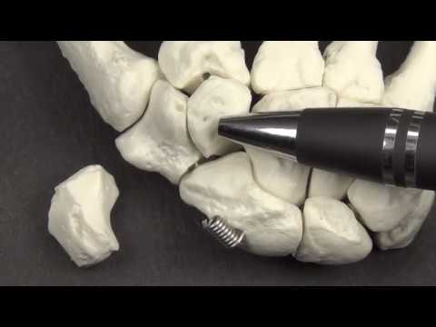
Trapezium Anatomy and Osteology with Side Determination

Показать описание
Mansoor Ahmed
This video reviews the anatomy and osteology of the trapezium, a left bone, and a right hand specimen are used.
Clinical Anatomy Course Director - Dr. Manion, PhD
Anatomy Lab Diener - Adam Jansen
1. Trapeziometacarpal joint- capable of opposition, articular surfaces are twisted, concavo-convex, and interlocking
2. Joint Stability - beak of the first metacarpal, trapezium recess, and ligaments
3. Ligaments - dorsal ligament complex, the volar beak ligament, and the intermetacarpal ligament
4. Palmar surface - tubercle, or ridge and a deep groove, flexor retinaculum (transverse carpal ligament), tendon of the flexor carpi radialis, superficial head of the flexor pollicis brevis, abductor pollicis brevis, opponens pollicis
5. Dorsal surface - groove for the radial artery
6. Lateral surface - radial collateral ligament
7. Proximal surface - scaphoid facet
8. Medial Surface - trapezoid facet
9. Distomedial Surface - facet for 2nd metacarpal
10. Blood Supply - radial artery, and branches of the dorsal and palmar carpal arch, extra osseous and intra-osseous blood supply
 0:06:04
 0:05:00
 0:01:07
 0:14:27
 0:02:37
 0:02:42
 0:09:07
 0:03:11
 0:02:26
 0:00:55
 0:02:25
 0:07:49
 0:00:06
 0:21:19
 0:00:27
 0:05:56
 0:14:33
 0:00:23
 0:10:42
 0:05:22
 0:00:30
 0:06:09
 0:12:53
 0:09:41