filmov
tv
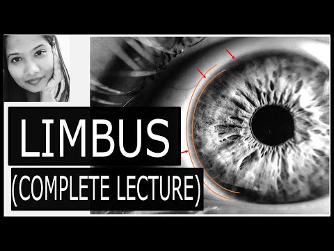
Anatomy of the Limbus (complete lecture) – Surgical Limbus, Incision site, Limbal Stem cells |

Показать описание
This video explains the anatomy of the limbus/corneoscleral junction from all perspectives- Histological, Anatomical & Surgical. It also explains the blue-grey zone & its significance while performing cataract surgeries, particularly during incision construction.
The diagrams shown in this video have been made as representative diagrams for the sole purpose of simplifying the topic. They should not be reproduced in either theory or viva exams.
This video also discusses limbal stem cells in detail – including palisades of Vogt & XYZ hypothesis. The understanding of limbal stem cell anatomy is important in order to understand various limbal stem cell diseases.
We also discuss a bit on the limbal vasculature.
I hope that medical undergraduate students, ophthalmology residents, nursing students, optometrists & practising ophthalmologists will find this video useful.
Please give your feedback in the comment section below.
Please like & share this video with your friends if you found it useful.
Please subscribe to my channel if you have an interest in ophthalmology & medicine, in general.
Thank you very much!
--------------- CHAPTERS------------------
0.00 - Intro
0.22 - Gross Anantomy
2.09 - Histological Limbus
6.17 - Transition occurring at the limbus
6.49 - Anatomical Limbus
8.22 - Surgical Limbus/ Blue Grey Zone
10.43 - Planning the surgical incision
11.38 - Limbal stem ccells
14.47 - Thoft’s XYZ hypothesis
15.22 - Limbal vasculature
15.41 - Significance of the Limbus
#Limbus #ophthalmology #anatomy
 0:16:36
Anatomy of the Limbus (complete lecture) – Surgical Limbus, Incision site, Limbal Stem cells |
 0:36:36
Anatomy Of Limbus Of Eye
 0:10:12
Anatomical Limbus
 0:16:07
Anatomy of the limbus complete lecture
 0:18:03
Anatomy Of Limbus - Dr. Zain Khatib
 0:05:16
LIMBUS ANATOMY
 0:03:18
|| Anatomy of Limbus || Histology of Limbus || #eye || #limbus
 0:01:48
Anatomy of limbus- Briefly explained...
 0:01:21
Exact Location for Surgical Limbus: The ideal site for Corneal Incision
 0:11:25
Limbus
 0:07:35
anatomy of limbus || limbus eye anatomy ||Session:4 ||2020||
 0:16:27
Limbus 1/5 - (General Information and Functions) || Eye Anatomy
 0:05:58
The Limbus Histology - Corneoscleral Junction | Med Madness
 0:27:01
Anatomy of Cornea , Sclera & Limbus | outer layers of eye | doctor Ron
 0:04:28
Limbus and its anatomy
 0:10:06
Cornea, sclera and limbus - eye histology part 1
 0:09:23
Limbus Anatomy 3/5 - Diagram || Eye Anatomy
 0:16:10
Eyeball - Outer Tunic Of Eyeball - Sclera |Cornea |Sceleo-Corneal Junction |Limbus |Canal Of Schlemm
 0:09:18
Limbus Anatomy in hindi
 0:00:53
cornea Limbus sclera 👁️👁️👁️👁️👁️🖊️🖊️🖊️🖊️👓👓👓📃📃📃⚔️⚔️⚔️...
 0:17:31
Limbus 5/5 - Histology & Anatomy by PPT Slide Share Images 🔥
 0:23:51
Cornea - Anatomy (Complete Lecture)|Layers,Hydration,Transparency,Blood & Nerve supply|Ophthalmo...
 0:01:02
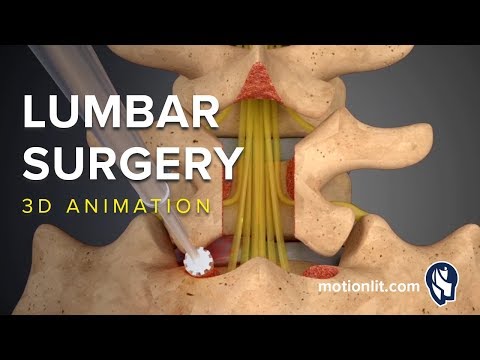
Lumbar Surgery - Laminectomy - 3D Medical Animation
 0:22:52
Eye- cornea -sclera- Limbus
Комментарии