filmov
tv
Histology of CEREBELLAR CORTEX

Показать описание
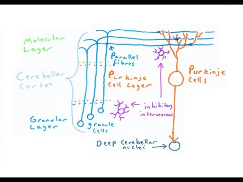
The grey matter of the cerebellum is also referred to as the cortex and may be split into three layers; the outer molecular layer, the middle layer of Purkinje cells and the inner granular layer. There are many neurons, glial cells and fibers located in the cortex which all contribute to the motor functions of the cerebellum.
Molecular layer
The outer molecular layer is synaptic and therefore contains many axons of granule cells and dendrites of the Purkinje cells with least density of cells. Superficially located stellate cells and basket cells are found in this layer. The stellate cells usually bear short dendrites in which make contact with small number of Purkinje cell dendrites. In comparison, basket cells have extensive dendritic processes that can make contact with much larger number of Purkinje cells. Both cells receive excitatory input from the parallel fibers and in turn exhibit inhibitory influence on the Purkinje cells
Purkinje cell layer
The middle layer (Purkinje cell layer) consists of a single layer of large pear-shaped Purkinje cells. Their cell bodies are largest in the cerebellum with unique and distinct appearance. The dendrites of these cells reside in the molecular layer, while their axons project deep through the granular layer and synapse into the deep nuclei of cerebellum.
Granular layer
Basket cell of cerebellum (Neuron cobiforme); Image:
Basket cell of cerebellum (Neuron cobiforme)
The inner granular layer contains many, tightly packed granule cells and Golgi type II cells. Granule cells, which are among the smallest neurons in the brain almost 5μm in diameter with round to oval in shape, usually represent the extensions of the mossy fibers. Their axons extend into the outer molecular layer where they branch in T shape forming parallel fibers and synapse with the dendrites of Purkinje, basket and stellate cells.
The nuclei of these granule cells generally stain dark, giving the whole granular layer a darker appearance compared to the white matter and molecular layer of the cortex. Golgi cells are also scattered throughout the granular layer, with their dendrites branching out in the molecular layer, while their axons synapses with the granule cells.
Mnemonic
There are a couple of mnemonics related to the cerebellar cortex histology.
First up, you can easily remember the three layers of the cerebellar cortex by using a mnemonic. ' MPG' or 'Mother Please Go' stands for:
Molecular
Purkinje
Granular
To remember the types of neurons present in the cerebellar cortex you can use the mnemonic ' Girls Bring Golden Stars in Pockets'. It stands for:
Golgi cells
Basket cells
Granular cells
Stellate cells
Purkinje cells
Cerebellar medulla
The inner medulla of white matter does not contain any cell bodies and therefore will stain a lighter colour in comparison to the grey matter cortex. It contains nerve fibers, supporting neuroglial cells and small blood vessels.
 0:08:04
Histology of CEREBELLAR CORTEX
 0:10:29
Cerebellar Histology - A Balanced Approach to Layers
 0:09:59
The Cerebellum
 0:06:18
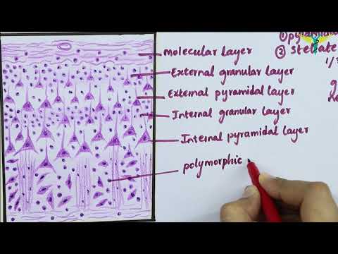
Histology of Cerebral Cortex.
 0:04:31
Cerebral Cortex Layers
 0:07:51
Central nervous system: Histology
 0:57:43
3-Histology of Cerebrum and Cerebellum-CNS
 0:03:27
Cerebral cortex (preview) - Human histology | Kenhub
 0:02:38
Cerebellum (H&E Staining) - PUMS Histology Slides Review Series
 0:08:24
Shotgun Histology Brain
 0:13:43
Glomerulus of the Cerebellum: A Histology Overview
 0:02:11
CEREBELLAR CORTEX |HISTOLOGY | Identifications Points
 1:03:56
Neurology | Cerebellum Anatomy & Function
 0:01:03
Cerebellar Cortex Made Easy! 🧠✨
 0:26:36
Histology of the brain
 0:05:26
SLIDE OF CEREBELLUM : HISTOLOGY
 0:00:28
cerebellum histology slide #cerebellum #histologia #histology #medical #anatomy #anatomymcq #ana
 0:07:39
Cerebellum Cortex Histology and Connections
 0:02:00
2-Minute Neuroscience: Cerebral Cortex
 0:05:07
The Six Layers of the Cerebral Cortex | Neuroscience 101
 0:02:45
Histology of cerebellum (practical)
 0:26:34
Neurology | Cerebral Cortex Anatomy & Function: Overview
 0:00:20
This Is What Connects Both Sides of Your Brain | The Corpus Callosum
 0:07:56
Histology with Chimmalgi: Cerebellum
Комментарии