filmov
tv
🧠 Tornwaldt Cyst on MRI 😱 What Radiologists MUST Know!

Показать описание
🧠 What is a Tornwaldt Cyst and how does it appear on MRI?
In this video, Dr. Martin, expert in head and neck radiology, explains the imaging features, clinical relevance, and differential diagnosis of the Tornwaldt cyst, a congenital nasopharyngeal lesion often found incidentally. Learn how to identify it on MRI and CT, when it becomes symptomatic, and how to differentiate it from more serious pathologies like nasopharyngeal carcinoma.
📍 Perfect for radiologists, ENT specialists, residents, and medical students preparing for exams or clinical practice.
🔍 Don’t forget to check the comment section for the Haller Cells video link — another must-know anatomical variant in head and neck imaging!
📌 Subscribe to Dr Martin: Head and Neck Radiology for more high-yield radiology content, anatomy tips, and real clinical cases.
📆 New videos every week!
 0:01:16
How to identify Thornwaldt Cyst on MRI
 0:03:56
🧠 Tornwaldt Cyst on MRI 😱 What Radiologists MUST Know!
 0:00:15
thornwaldt cyst
 0:01:01
Thornwaldt cyst at upper nasopharyngeal mucosa. #mri #ct #viralvideo #viral #shorts
 0:00:14
Thornwaldts cyst in a ct scan dr sbv
 0:00:31
GET RELIEF FROM TORNWALDT CYST WITH HOMEOPATHY
 0:00:16
Not surprised
 0:00:55
Découverte fortuite d'une masse nasopharyngée dans un Kyste de Tornwaldt
 0:33:41
'IMAGING CYSTIC NECK MASSES Dr /Ahmed Esawy
 0:04:16
Nasolabial cyst. Malayalam.Patient teaching programme.
 1:51:27
CYST CAUSES SIGNS SYMPTOMS DIAGNOSIS MANAGEMENT.
 0:01:47
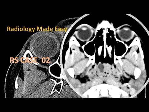
A Patient with history of head trauma | FRCR RS Cases 01
 0:51:25
Cervical Spine Masses | Pt. 1
 0:06:29
EXPERT DDX BRAIN AND SPINE IMAGING
 0:03:04
CT & MRI lesion Discussion case 1 to 7
 0:30:12
Spaces of Neck
 0:00:47
REMOVAL OF NASAL CYST - DR. TANVEER JANJUA
 0:46:30
Imaging of Skull base lesions
 1:57:18
7 radiology channel imaging oral Board of neck imaging
 0:50:02
HEAD AND NECK RADIOLOGY CASE REVIEW SERIES
 1:28:25
HEAD AND NECK EXPERT DDX
 0:47:10
Imaging of nasopharynx
 0:51:37
Neck Film ReadingNasopharynx Lecture 111
 0:25:03
HEAD AND NECK DIAGNISTIC IMAGING SERIES
Комментарии