filmov
tv
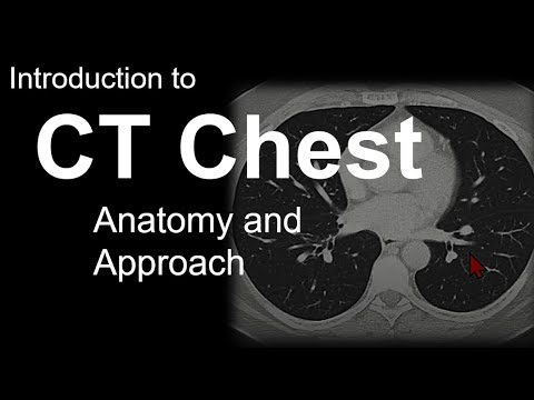
[CT] Chest | Search Pattern

Показать описание
[2:15] - A quick scroll through axial images can give you a sense of overall limitations/artifact and distribution of pathology.
[4:30] - Be careful of nodules near the hilum and lung bases. These are common blind spots on CT.
[5:27] - On contrast enhanced exams, look for filling defects/clots, especially at the left trial appendage, posterior left atrial wall, left ventricular apex, and any area with evidence of prior infarct.
“Search Pattern” is a video series designed to provide an accessible introduction to evaluating diagnostic imaging in a systematic fashion.
Disclaimer: This is an introduction to study interpretation. An enormous amount of detail is omitted for brevity. Continue dedicated reading, seeing as many cases as possible, and getting feedback from subspecialists during the course of your training.
Long Tu, MD’s book “Search Pattern: A Systematic Approach to Diagnostic Imaging” can be purchased at this link:
![[CT] Chest |](https://i.ytimg.com/vi/lnNG_s1EidM/hqdefault.jpg) 0:11:42
0:11:42
 0:17:21
 0:36:54
 0:11:18
 0:26:20
 0:22:53
 0:12:26
![[NM] PET CT](https://i.ytimg.com/vi/VsspJQVPmpc/hqdefault.jpg) 0:13:21
0:13:21
 0:12:42
 0:13:34
 0:16:25
 0:06:43
![[CT] CTA for](https://i.ytimg.com/vi/cmdU148Rnn4/hqdefault.jpg) 0:07:04
0:07:04
 0:12:41
 0:00:15
 0:03:40
 1:05:32
 0:19:40
 0:03:43
![[XR] Chest Radiograph](https://i.ytimg.com/vi/o0jIbCQGXSo/hqdefault.jpg) 0:08:32
0:08:32
 0:13:58
 0:11:09
 0:07:00
 0:09:05