filmov
tv
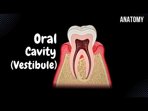
Oral Vestibule (Lips, Cheeks, Teeth, Gums) - Oral Cavity Anatomy

Показать описание
Content:
0:00 Introduction
1:57 External Structures of the Mouth
2:49 Division of the Oral Cavity
3:02 Oral Vestibule
3:24 Anatomy of the Lips
4:07 Anatomy of the Cheeks
5:28 Anatomy of the Teeth
8:24 Tooth Arrangement
10:15 Anatomy of the Gums
-------------------------------
-------------------------------
General structures of the digestive system:
- Oral Cavity
- Pharynx
- Oesophagus
- Stomach
- Small intestine
- Large Intestine
Accessory Organs
- Teeth
- Tongue
- Salivary glands
- Liver
- Pancreas
- Gall Bladder
External Structures of the Mouth:
- Upper Lip (Labium Superior)
- Lower Lip (Labium Inferior)
- Oral Angle (Labial Commissure)
- Nasolabial Sulcus (Sulcus Nasolabialis)
- Philtrum
- Mentolabial Sulcus (Sulcus Mentolabialis)
- Oral Fissure (Rima Oris)
Division of the Oral Cavity:
- Oral Vesitibule (Vestibulum Oris)
- Oral Cavity Proper (Cavitas Oris Propria)
Oral Vestibule:
- External Borders: Lips and Cheeks
- Internal Borders: Teeth and Gums
Anatomy of the Lips:
- Frenulum of the Upper Lip (Frenulum Labii Superioris)
- Frenulum of the Lower Lip (Frenulum Labii Inferioris)
- Labial Glands (Glandulae Labialis)
Anatomy of the Cheeks:
- Buccinator Muscle (musculus buccinator)
- Buccopharyngeal Fascia
- Buccal Fat Pad (Bichat's Fat Pad)
- Layers of the Skin (cutis)
- Tunica Mucosa
- Parotid Duct (Ductus Parotideus)
- Papilla of the Parotid Duct (Papillae Ductus Parotidei)
Anatomy of the Teeth:
- Crown of the tooth (Corona Dentis)
- Root of the tooth (Radix Dentis)
- Neck of the tooth (Cervix Dentis)
- Periodontium
- Gomphosis (Dentoalveolar Joint)
- Dental Pulp (Pulpa Dentis)
- Root Canal (Canalis Radicis Dentis)
- Apical Foramen (Foramen Apicis Dentis)
- Dentin (Dentinum)
- Enamel (Enamelum)
- Cement (Cementum)
- Milk Teeth (Dentes decidui) 20 teeth, begins at 6th moth of life and ends at 24th month of life
- Replacement of milk teeth at age of 6-12 years
- Permanent Teeth (Dentes Permanentes) 32 teeth
- Wisdom teeth (dentes serotinus) appears at the age of 17-24 years
Tooth Arrangement:
PS! European way of arrangement
- First we divide the teeth into 4 equal quadrants
- First 2 teeth are called Incisor Teeth (Dentes Invisivi)
- Next teeth are Canine Teeth (Dentes Canini)
- Premolar Teeth (Dentes Premolares)
- Molar Teeth (Dentes Molares)
- 8*4=32 means we have 32 premanent teeth
- Milk Teeth have two incisor teeth, one canine tooth and two molar teeth
Anatomy of the Gums (Gingiva):
- Alveolar Mucus Membrane (Covers at the level of root of the tooth. Contains a lot of submucosal Connective tissue)
- Gum Proper (attached to the periosteum)
- Gingival Papillae (papillae gingivales)
- Gingival Margin (margo gingivalis)
- Gingival Sulcus (sulcus gingivalis)
Sources used in this video:
- Memorix Anatomy 2nd Edition by Hudák Radovan (Author), Kachlík David (Author), Volný Ondřej (Author)
- Biorender
- University notes and lectures
 0:12:07
Oral Vestibule (Lips, Cheeks, Teeth, Gums) - Oral Cavity Anatomy
 0:14:59
Oral cavity anatomy
 0:02:01
Overview of the Oral Cavity (preview) - Human Anatomy | Kenhub
 0:22:38
Oral cavity | Oral vestibule (lips,teeth,cheeks)| Digestive system | splancology
 0:07:43
Complete Oral Cavity Anatomy | Vestibule, Teeth, Gums, Lips, Cheeks & More
 0:05:31
anatomy of digestive system-ORAL CAVITY,VESTIBULE AND LIPS
 0:11:10
ORAL CAVITY ANATOMY ( VESTIBULE OF MOUTH)
 0:10:08
Anatomy of oral cavity| vestibule anatomy| anatomy of lips, Cheeks and teeth
 4:42:34
Don’t Watch This If You’re Tired — Rest & Relaxation (Video Book)
 0:00:19
Parts of the Mouth #viral #fyp #learnenglish
 0:16:14
Oral Cavity Proper (Palate & Tongue) - Oral Cavity Anatomy
 0:13:34
ANATOMY OF MOUTH
 0:15:26
Oral cavity & oral Vestibule | Dissection room 1 | Anatomy
 0:16:30
Blood and nerve supply of the oral cavity
 0:07:58
Anatomy and Physiology: Oral Cavity
 0:00:59
Oral Cavity in 1 min
 0:22:01
Anatomy of the Oral Cavity, Dr Adel Bondok Making Anatomy Easy
 0:09:39
oral cavity (vestibule, lips , cheeks)
 0:03:00
Bump inside mouth - Parotid duct Diseases. Stensen duct; Sialolithiasis
 0:07:07
Oral Cavity Boundaries and Contents
 0:07:58
Anatomy and Physiology Oral Cavity
 0:01:13
Oral Cavity Anatomy, Functions, and Dental Structure Explained
 0:04:38
Digestion in the Mouth
 0:00:11
Parts of Human mouth
Комментарии