filmov
tv
Optical Coherence Tomography - OCT (Full)

Показать описание
INTRODUCTION:
-------------------------
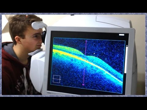
• OCT is an optical instrument that can perform cross-sectional image of biological tissue within less than 10 micron axial resolution using light waves
• Retina is easily accessible to the external light, hence it is specially suited for retinal disorder
• The information provided by OCT is similar to in vivo histopathology of the retina
EXAMPLE:
Zeiss stratus OCT
Topcon 3D OCT-1000
PRINCIPLE OF OCT:
--------------------------------
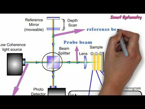
• It is a imaging technology projected light beam (820nm) near infrared light
• The beam is then split into two beam (Probe beam & Reference beam) by Beam splitter
• Probe beam reach to the target tissue (retina) & reference beam reach to the reference mirror
at a known distance
• The echo time delay of light reflected various layer of target tissue (retina) is compared with
the echo time delay of light reflected from the reference mirror
• A positive interference is produced when light reflected from target tissue & reference mirror
arrives simultaneously
• This interference is measured by a photodetector which finally produce a range of time delays
for comparison
• The interferometer integrates several data points over 2mm depth to construct a tomogram
of retinal structures
• It is real time tomogram using false color scale & different colors represent light
backscattering from the different layers of retina
OCT SYSTEM CONSIST OF:
------------------------------------------
• Fundus viewing unit
• Interferometer unit
• Computer display
• Control panel
• Color inkjet printer
GENERATION OF
----------------------------
• OCT- 1:
o 1st generation
o Transverse resolution 20 micron
o Axial resolution 10 micron
• OCT-2:
o 2nd generation
o Transverse resolution 20 micron
o Axial resolution 10 micron
o Better user interference
• BOTH OCT-1 & OCT-2:
o acquire 100 vertical scan in approximately 1.2 sec
• OCT-3:
o 3rd generation
o Axial resolution 7-8 micron
o Acquire 512 vertical scan
COLOR CODING OF OCT SCAN:
------------------------------------------------
• RED-YELLOW COLORS: represents areas of maximal optical reflection & backscattering
• BLUE & BLACK: Represents areas of minimal optical refelection & backscattering
VARIOUS PATTERN OF B-SCAN:
--------------------------------------------------
• CIRCULAR SCAN FOR THE ONH RNFL:
This generates a plot of the peripapillary RNFL thickness which is important in
glaucoma diagnosis & monitoring
• RADIAL LINE THROUGH ONH:
Consist of 6-24 slices through a common central point on the ONH
• MACULAR RADIAL LINES: Used to measure retinal thickness
PROCEDURE OF OCT:
"""""""""""""""""""""""""""""""""""
• STEP-1: PATIENTS DATA
Activation of instrument & entering patients data
• STEP-2: PATIENTS PREPARATION:
Pupil dilate with mydriatics (tropicamide)
Asked to look into the internal fixation target light in the
ocular lens
• STEP-3: PROTOCOL FOR SCAN ACQUISITION:
Selected as per the case requirements
The scanning beam is placed on the target area and scans
are obtained
• STEP-4: PRODUCTION & DISPLAY IMAGE:
Several data points are integrated by the interferometer to
construct a tomogram of the target area
The tomogram is displayed in either grey scale or false color
on a high-resolution computer screen
-------------------------
• OCT is an optical instrument that can perform cross-sectional image of biological tissue within less than 10 micron axial resolution using light waves
• Retina is easily accessible to the external light, hence it is specially suited for retinal disorder
• The information provided by OCT is similar to in vivo histopathology of the retina
EXAMPLE:
Zeiss stratus OCT
Topcon 3D OCT-1000
PRINCIPLE OF OCT:
--------------------------------
• It is a imaging technology projected light beam (820nm) near infrared light
• The beam is then split into two beam (Probe beam & Reference beam) by Beam splitter
• Probe beam reach to the target tissue (retina) & reference beam reach to the reference mirror
at a known distance
• The echo time delay of light reflected various layer of target tissue (retina) is compared with
the echo time delay of light reflected from the reference mirror
• A positive interference is produced when light reflected from target tissue & reference mirror
arrives simultaneously
• This interference is measured by a photodetector which finally produce a range of time delays
for comparison
• The interferometer integrates several data points over 2mm depth to construct a tomogram
of retinal structures
• It is real time tomogram using false color scale & different colors represent light
backscattering from the different layers of retina
OCT SYSTEM CONSIST OF:
------------------------------------------
• Fundus viewing unit
• Interferometer unit
• Computer display
• Control panel
• Color inkjet printer
GENERATION OF
----------------------------
• OCT- 1:
o 1st generation
o Transverse resolution 20 micron
o Axial resolution 10 micron
• OCT-2:
o 2nd generation
o Transverse resolution 20 micron
o Axial resolution 10 micron
o Better user interference
• BOTH OCT-1 & OCT-2:
o acquire 100 vertical scan in approximately 1.2 sec
• OCT-3:
o 3rd generation
o Axial resolution 7-8 micron
o Acquire 512 vertical scan
COLOR CODING OF OCT SCAN:
------------------------------------------------
• RED-YELLOW COLORS: represents areas of maximal optical reflection & backscattering
• BLUE & BLACK: Represents areas of minimal optical refelection & backscattering
VARIOUS PATTERN OF B-SCAN:
--------------------------------------------------
• CIRCULAR SCAN FOR THE ONH RNFL:
This generates a plot of the peripapillary RNFL thickness which is important in
glaucoma diagnosis & monitoring
• RADIAL LINE THROUGH ONH:
Consist of 6-24 slices through a common central point on the ONH
• MACULAR RADIAL LINES: Used to measure retinal thickness
PROCEDURE OF OCT:
"""""""""""""""""""""""""""""""""""
• STEP-1: PATIENTS DATA
Activation of instrument & entering patients data
• STEP-2: PATIENTS PREPARATION:
Pupil dilate with mydriatics (tropicamide)
Asked to look into the internal fixation target light in the
ocular lens
• STEP-3: PROTOCOL FOR SCAN ACQUISITION:
Selected as per the case requirements
The scanning beam is placed on the target area and scans
are obtained
• STEP-4: PRODUCTION & DISPLAY IMAGE:
Several data points are integrated by the interferometer to
construct a tomogram of the target area
The tomogram is displayed in either grey scale or false color
on a high-resolution computer screen
 0:03:27
Optical Coherence Tomography: A new way of seeing
 0:03:46
What is OCT Scanning? (Optical Coherence Tomography)
 0:11:09
Optical Coherence Tomography - OCT (Full)
 0:01:51
Optical Coherence Tomography (OCT) in MS
 0:49:17
Optical Coherence Tomography Imaging Analysis for Retinal Disease
 0:01:13
What is OCT? How Optical Coherence Tomography Works I Retinal disease evaluation tool
 0:31:32
WHAT IS OCT || OCT technology explained ||
 0:04:35
Optical Coherence Tomography (OCT) Full System Assembly
 0:04:38
The Invention of Optical Coherence Tomography
 0:00:31
What is an OCT test?
 0:06:18
Optical Coherence Tomography - OCT | part-1
 0:05:52
OCT Angiography: Optical Coherence Tomography (OCT) Technology for Coronary Imaging in Angioplasty
 0:02:13
Optical coherence tomography (OCT)
 0:03:12
Optical Coherence Tomography (OCT)
 0:00:52
NIH SPARC Program Tools & Tech: Optical coherence tomography (OCT) (SPARC Plug: Tools & Tech...
 0:00:42
Optical Coherence Tomography (OCT) | Learn From Expert | Medipulse Hospital
 0:01:00
High-precision tiniest Endoscopic Optical Coherence Tomography (OCT)
 0:04:33
OCT SCAN | Optical Coherence Tomography | Retina
 0:03:23
Optical Coherence Tomography(OCT) in Interventional Cardiology
 0:12:07
Julia Walther: Optical coherence tomography in the oral cavity
 0:00:14
What is an OCT?
 0:03:40
OCT Scan | All you need to know about Optical Coherence Tomography | Specsavers UK & ROI
 0:00:16
OCT scan of the retina takes just a few seconds to give incredible details of the retina.
 0:30:42
OCT (Optical Coherence Tomography)- Elias Hanna, Univ of Iowa
Комментарии