filmov
tv
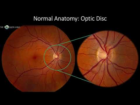
Imaging of the optic nerve

Показать описание
Imaging of the eye, orbits and visual tracts (part 4): the conal space.
This is the fourth video in a series on orbital imaging. In this video we talk about lesions located in the intraconal compartment. The intraconal compartment contains the optic nerve, so a lot of this presentation will focus on optic nerve pathology.
This video is brought to you by the neuroradiologist:
#radiology #neuroradiology #neurology #medicalstudent #neuroradiologist #theneuroradiologist #MRI #CT #medical #eyes #mri #radiologytechnologist #radiologyresident #brainanatomy #ophthalmology #orbit
 0:26:45
Imaging of the optic nerve
 0:03:59
Understanding Optic Nerve OCT
 0:16:37
OPTIC NERVE pathologies- SIMPLIFIED
 0:35:51
Lecture: Examining the Optic Nerve
 0:20:11
Cranial Nerve Anatomy on MRI
 0:49:43
Optic Nerve Pathology
 0:02:38
Optic Nerve Sheath Meningioma
 0:04:30
Optic Nerve Glioma. Easy to understand on Radiology MR images
 0:39:31
An Introduction to Advanced MRI techniques: fMRI, spectroscopy, perfusion & diffusion tensor ima...
 0:16:51
Ocular and Optic Nerve Trauma | Free Radiology CME
 0:03:45
Optic nerve tumors
 0:01:10
Optic neuritis (Radiopaedia.org) Cases in Radiology
 0:09:01
RADIOLOGY OF OPTIC PATHWAY GLIOMA
 0:01:00
Normal Disc vs. Glaucomatous Disc vs. Atrophic Disc #Basics #Shorts
 0:00:57
Optic Atrophy - Clinical Appearance of Each Type #Shorts
 0:01:18
Taking an Optic Nerve Photo | Dr. Alan Mendelsohn
 0:04:55
Optic Atrophy Explained: Causes, Symptoms, and Treatment Options
 0:05:42
Fundus Photography Interpretation
 0:01:33
IIH: MRI Showing Flattening of Posterior Globes and Distal Optic Nerve Sheath Dilatation
 0:00:05
Adaptive optics retinal imaging - Optic Nerve Head
 0:00:22
Optic nerve head analysis
 0:05:48
IIH: MRI Findings in a Patient with Blurry Vision, Headaches, Tinnitus and Papilledema on Fundoscopy
 0:10:39
Optic nerve head microarchitecture: en-face OCT and OCT angiographic imaging of lamina cribrosa,
 0:56:53
Orbital and Neuro-Ophthalmological Imaging
Комментарии