filmov
tv
NORMAL MACULAR ANATOMY ON OCT

Показать описание
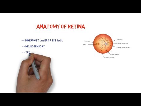
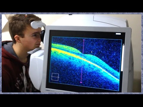
This video talks about the normal macular and retinal anatomy on OCT scan. the normal macular anatomy on oct can be studied by noticing the vitreo-retinal interface, the inner retinal layer, the outer retinal layer and the choroid and the sclera . The VR interface is the junction between the vtreous and the ILM ( internal limiting memberane of the retina. The inner retina consists of layers of hyperreflective and hyporeflective zones on OCT. the Outer retinal layer is again divided into 4 parts ( the outer limiting memberane, ellipsoid zone,interdigitation zone, the RPE-Bruchs comples. Using the enhanced depth imaging, the choroid and retina can also be images and studies on OCT
#oct
#ophthalmology
#retina
#oct
#ophthalmology
#retina
 0:18:06
NORMAL MACULAR ANATOMY ON OCT
 0:13:53
HOW TO READ MACULAR OCT PRINTOUT? made easy!!
 0:02:04
Macular anatomy - basic layers on OCT
 0:06:47
OCT Tutorial On Macular Anatomy part 1
 0:01:00
Normal Disc vs. Glaucomatous Disc vs. Atrophic Disc #Basics #Shorts
 0:50:07
Macular OCT Interpretation: A Practical Discussion with Dr. David E. Lederer
 0:56:58
Ordering and Interpreting Retinal OCT Images
 0:00:56
Fundus of an EYE explained simply in less than 60 seconds ! USMLE, NEET PG, NCLEX
 0:00:26
What is the Retina?
 0:00:22
Solar Retinopathy #eyedisease #ophthalmology #retina #oct
 0:05:00
Normal Macular OCT Line Scan
 0:23:38
HOW TO READ AN OCT PRINTOUT IN GLAUCOMA || BASIC TESTING PROTOCOLS|| ( RNFL, ONH & MACULAR ANALY...
 0:47:03
Qualitative analysis of OCT macula: OCT interpretation for macular pathologies
 0:04:13
Anatomy of Retina || mnemonic for 10 layers ||
 0:00:22
Optic nerve head analysis
 2:00:08
Mastering OCT Interpretation with Dr. Mark Friedberg
 0:03:59
Understanding Optic Nerve OCT
 0:05:42
Fundus Photography Interpretation
 0:00:53
Basic Eye Anatomy by Vicki Chan MD
 0:03:46
What is OCT Scanning? (Optical Coherence Tomography)
 0:00:59
Eye doctor explains: basic anatomy #eye #anatomy #youtubehealth
 0:15:17
OCT in AMD( Age related macular degeneration
 0:00:18
Retinal Arteriolar Macroaneurysm
 0:00:15
Human Eye Anatomy //Eye Anatomy//Eye #eyes #eye
Комментарии