filmov
tv
Radial Nerve Anatomy - Everything You Need To Know - Dr. Nabil Ebraheim
Показать описание
Dr. Ebraheim’s educational animated video describes the anatomy associated with the radial nerve.
Follow me on twitter:
Radial Nerve Anatomy
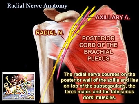
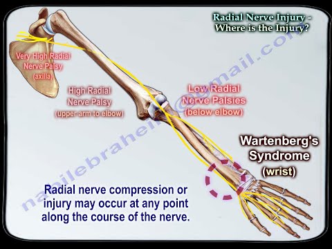
The radial nerve arises from the posterior cord of the brachial plexus and lies posterior to the axillary artery. The radial nerve receives branches from each nerve root from C5-T1. The radial nerve courses on the posterior wall of the axilla and lies on top of the subscapularis, the teres major, and the latissimus dorsi muscles. All three of these muscles (subscapularis-upper & lower subscapular n., teres major-lower subscapular n., latissimus dorsi-thoracodorsal n.) are supplied by the posterior cord of the brachial plexus. The radial nerve continues into the posterior compartment of the upper arm. The radial nerve then gives three branches in the axilla: branch to long head of triceps, branch to medial head of triceps, posterior cutaneous nerve of the arm. Some books show the position of the posterior cutaneous nerve of the arm may be higher than the branches to the triceps. The profunda brachii artery arises from the axillary artery. The radial nerve next travels through the triangular interval with the profunda brachii artery posteriorly. The radial nerve is bound proximally by the teres major, medially by the long head triceps, and laterally by the humeral shaft. It contains the profunda brachii artery and the radial nerve. The radial nerve enters the upper arm between the long head and the medial head of the triceps and then it runs towards the spiral groove of the humerus. The spiral groove is a thin, bare area of bone that lies in the upper 2/3 of the back of the humerus between the lateral and medial heads of the triceps. There are posterior safe zones of the humerus 10cm distal to the lateral acromion and 10cm proximal to the lateral epicondyle. Four branches arise from the radial nerve within the spiral groove. In the posterior approach, if the surgeon follows these cutaneous nerves proximally, it will lead to identification of the radial nerve itself. The radial nerve passes through the lateral intermuscular septum enters the anterior compartment of the arm above the elbow joint. Anteriorly, it runs between the brachialis and brachioradialis muscle anterior to the lateral epicondyle. This is the site for exposure of the radial nerve anteriorly (between the brachialis and brachioradialis muscles anteriorly). The radial nerve gives branches to supply the lateral part of the brachialis, brachioradialis, extensor carpi radialis longus, and the extensor carpi radialis brevis muscles. The radial nerve is vulnerable to injury below the spinal groove when there is a fracture in the distal third of the humeral shaft. Injury to the nerve will cause wrist drop. At about the level of the lateral epicondyle, the radial nerve begins to divide into the deep branch and the superficial branch of the radial nerve. The anconeus muscle is also innervated by the radial nerve. The posterior interosseous nerve (deep branch) enters the extensor compartment of the forearm between the two heads of the supinator muscle. The ulnar nerve enters the forearm by passing through the two heads of the flexor carpi ulnaris muscle and the median nerve enters the forearm by passing through the two heads of the pronator teres muscle. The area in which the posterior interosseous nerve passes through is called the “Arcade of Frohse” and this area is often a site of entrapment of the nerve. The posterior interosseous nerve supplies these muscles on the radial side and dorsal surface of the forearm: posterior interosseous n., posterior interosseous n. passes through the supinator m., extensor digiti minimi, extensor carpi ulnaris, extensor digitorum, extensor indicis, extensor pollicis brevis, extensor pollicis longus, and abductor pollicis longus. The posterior interosseous nerve does not supply cutaneous sensation and it is purely motor nerve. Injury to the posterior interosseous nerve will lead to inability of the patient to extend their fingers or “hitchhike” the thumb. During recovery from posterior interosseous nerve injury, the extensor digitorum muscle is the first one to recover and the extensor indicis is the last muscle to recover. The superficial radial nerve runs deep to the brachioradialis muscle. The superficial radial nerve continues until about 5cm above the wrist where it immerges from underneath the brachioradialis muscle, piercing the deep fascia and lying between the brachioradialis and the extensor carpi radialis longus muscles, then descending towards the anatomical snuff box. The superficial radial nerve is a sensory nerve supplying the majority of the dorsum of the hand. The sensory areas involving the cutaneous branches of the upper arm and forearm, and the superficial sensory radial nerve are shown here. Wartenberg’s Syndrome is characterized by entrapment of the superficial branch of the radial nerve above the wrist.
Follow me on twitter:
Radial Nerve Anatomy
The radial nerve arises from the posterior cord of the brachial plexus and lies posterior to the axillary artery. The radial nerve receives branches from each nerve root from C5-T1. The radial nerve courses on the posterior wall of the axilla and lies on top of the subscapularis, the teres major, and the latissimus dorsi muscles. All three of these muscles (subscapularis-upper & lower subscapular n., teres major-lower subscapular n., latissimus dorsi-thoracodorsal n.) are supplied by the posterior cord of the brachial plexus. The radial nerve continues into the posterior compartment of the upper arm. The radial nerve then gives three branches in the axilla: branch to long head of triceps, branch to medial head of triceps, posterior cutaneous nerve of the arm. Some books show the position of the posterior cutaneous nerve of the arm may be higher than the branches to the triceps. The profunda brachii artery arises from the axillary artery. The radial nerve next travels through the triangular interval with the profunda brachii artery posteriorly. The radial nerve is bound proximally by the teres major, medially by the long head triceps, and laterally by the humeral shaft. It contains the profunda brachii artery and the radial nerve. The radial nerve enters the upper arm between the long head and the medial head of the triceps and then it runs towards the spiral groove of the humerus. The spiral groove is a thin, bare area of bone that lies in the upper 2/3 of the back of the humerus between the lateral and medial heads of the triceps. There are posterior safe zones of the humerus 10cm distal to the lateral acromion and 10cm proximal to the lateral epicondyle. Four branches arise from the radial nerve within the spiral groove. In the posterior approach, if the surgeon follows these cutaneous nerves proximally, it will lead to identification of the radial nerve itself. The radial nerve passes through the lateral intermuscular septum enters the anterior compartment of the arm above the elbow joint. Anteriorly, it runs between the brachialis and brachioradialis muscle anterior to the lateral epicondyle. This is the site for exposure of the radial nerve anteriorly (between the brachialis and brachioradialis muscles anteriorly). The radial nerve gives branches to supply the lateral part of the brachialis, brachioradialis, extensor carpi radialis longus, and the extensor carpi radialis brevis muscles. The radial nerve is vulnerable to injury below the spinal groove when there is a fracture in the distal third of the humeral shaft. Injury to the nerve will cause wrist drop. At about the level of the lateral epicondyle, the radial nerve begins to divide into the deep branch and the superficial branch of the radial nerve. The anconeus muscle is also innervated by the radial nerve. The posterior interosseous nerve (deep branch) enters the extensor compartment of the forearm between the two heads of the supinator muscle. The ulnar nerve enters the forearm by passing through the two heads of the flexor carpi ulnaris muscle and the median nerve enters the forearm by passing through the two heads of the pronator teres muscle. The area in which the posterior interosseous nerve passes through is called the “Arcade of Frohse” and this area is often a site of entrapment of the nerve. The posterior interosseous nerve supplies these muscles on the radial side and dorsal surface of the forearm: posterior interosseous n., posterior interosseous n. passes through the supinator m., extensor digiti minimi, extensor carpi ulnaris, extensor digitorum, extensor indicis, extensor pollicis brevis, extensor pollicis longus, and abductor pollicis longus. The posterior interosseous nerve does not supply cutaneous sensation and it is purely motor nerve. Injury to the posterior interosseous nerve will lead to inability of the patient to extend their fingers or “hitchhike” the thumb. During recovery from posterior interosseous nerve injury, the extensor digitorum muscle is the first one to recover and the extensor indicis is the last muscle to recover. The superficial radial nerve runs deep to the brachioradialis muscle. The superficial radial nerve continues until about 5cm above the wrist where it immerges from underneath the brachioradialis muscle, piercing the deep fascia and lying between the brachioradialis and the extensor carpi radialis longus muscles, then descending towards the anatomical snuff box. The superficial radial nerve is a sensory nerve supplying the majority of the dorsum of the hand. The sensory areas involving the cutaneous branches of the upper arm and forearm, and the superficial sensory radial nerve are shown here. Wartenberg’s Syndrome is characterized by entrapment of the superficial branch of the radial nerve above the wrist.
Radial Nerve Anatomy - Everything You Need To Know - Dr. Nabil Ebraheim
 0:07:01
Radial Nerve | 3D Anatomy Tutorial
 0:02:35
Radial Nerve - Branches, Course & Innervation - Human Anatomy | Kenhub
 0:06:56
Radial Nerve anatomy - Everything You Need To Know - Dr. Nabil Ebraheim
 0:03:26
Course Of The Radial Nerve Simplified - Everything You Need To Know - Dr. Nabil Ebraheim
 0:04:03
Radial Nerve - part #1 | Anatomy Tutorial
 0:07:58
Radial Nerve Injury,Where Is The Injury - Everything You Need To Know - Dr. Nabil Ebraheim
 0:03:13
1. UL Radial Nerve - Course
 0:09:52
RADIAL NERVE | ANATOMY | SIMPLIFIED
 0:37:34
Radial nerve anatomy : Course and branches.
 0:00:35
Radial Nerve : muscle supply - mnemonic | #shorts
 0:18:05
Ulnar, Median and Radial Nerves, Dr Adel Bondok
 0:07:43
Radial Nerve Overview | Branches & Functions
 0:29:29
Radial Nerve Anatomy | Upper Limb Anatomy
 0:04:54
Radial Nerve Palsy, injury - WRIST DROP . Everything You Need To Know - Dr. Nabil Ebraheim
 0:04:29
Radial Nerve Entrapment Release - MSR Protocol - Part 4
 0:26:56
Nerves of the upper limb (anatomy)
 0:00:59
Shorts #12: Branches of the Radial Nerve - In less than a minute!
 0:00:16
radial nerve......short notes hand written notes 👍 anatomy 🥰🖊️
 0:17:21
Radial Nerve
 0:10:46
Radial nerve anatomy in hindi | Radial nerve 3D Anatomy | Radial nerve branches anatomy
 0:03:59
Quick & Easy Upper Limb Nerve Tests | Radial, Median, Ulna, AIN Nerves: Rock, Paper, Scissors, O...
 0:29:09
RADIAL NERVE ULTRASOUND
 0:02:53
wrist drop anatomy radial nerve clinical anatomy | clinical anatomy of radial nerve wrist drop
Комментарии