filmov
tv
Molecular Mechanism of Gram Staining

Показать описание
Gram stain or Gram staining, also called Gram's method, is a method of staining used to distinguish and classify bacterial species into two large groups (gram-positive and gram-negative). The name comes from the Danish bacteriologist Hans Christian Gram, who developed the technique.
Gram staining differentiates bacteria by the chemical and physical properties of their cell walls by detecting peptidoglycan, which is present in the cell wall of Gram-positive bacteria. Gram-negative cells also contain peptidoglycan, but a very small layer of it that is dissolved when the alcohol is added. This is why the cell loses its initial colour from the primary stain.[1] Gram-positive bacteria retain the crystal violet dye, and thus are stained violet, while the Gram-negative bacteria do not; after washing, a counterstain is added (commonly safranin or fuchsine) that will stain these Gram-negative bacteria a pink color. Both Gram-positive bacteria and Gram-negative bacteria pick up the counterstain. The counterstain, however, is unseen on Gram-positive bacteria because of the darker crystal violet stain.
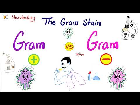
The Gram stain is almost always the first step in the preliminary identification of a bacterial organism. While Gram staining is a valuable diagnostic tool in both clinical and research settings, not all bacteria can be definitively classified by this technique. This gives rise to gram-variable and gram-indeterminate groups.
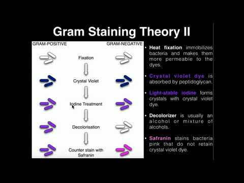
Gram-positive bacteria have a thick mesh-like cell wall made of peptidoglycan (50–90% of cell envelope), and as a result are stained purple by crystal violet, whereas gram-negative bacteria have a thinner layer (10% of cell envelope), so do not retain the purple stain and are counter-stained pink by safranin. There are four basic steps of the Gram stain:
Applying a primary stain (crystal violet) to a heat-fixed smear of a bacterial culture. Heat fixation kills some bacteria but is mostly used to affix the bacteria to the slide so that they don't rinse out during the staining procedure
The addition of iodide, which binds to crystal violet and traps it in the cell
Rapid decolorization with ethanol or acetone
Counterstaining with safranin. Carbol fuchsin is sometimes substituted for safranin since it more intensely stains anaerobic bacteria, but it is less commonly used as a counterstain.
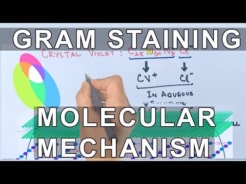
Crystal violet (CV) dissociates in aqueous solutions into CV+
and chloride (Cl−
) ions. These ions penetrate through the cell wall and cell membrane of both gram-positive and gram-negative cells. The CV+
ion interacts with negatively charged components of bacterial cells and stains the cells purple.[11]
Iodide (I−
or I−
3) interacts with CV+
and forms large complexes of crystal violet and iodine (CV–I) within the inner and outer layers of the cell. Iodine is often referred to as a mordant, but is a trapping agent that prevents the removal of the CV–I complex and, therefore, color the cell.
Gram staining differentiates bacteria by the chemical and physical properties of their cell walls by detecting peptidoglycan, which is present in the cell wall of Gram-positive bacteria. Gram-negative cells also contain peptidoglycan, but a very small layer of it that is dissolved when the alcohol is added. This is why the cell loses its initial colour from the primary stain.[1] Gram-positive bacteria retain the crystal violet dye, and thus are stained violet, while the Gram-negative bacteria do not; after washing, a counterstain is added (commonly safranin or fuchsine) that will stain these Gram-negative bacteria a pink color. Both Gram-positive bacteria and Gram-negative bacteria pick up the counterstain. The counterstain, however, is unseen on Gram-positive bacteria because of the darker crystal violet stain.
The Gram stain is almost always the first step in the preliminary identification of a bacterial organism. While Gram staining is a valuable diagnostic tool in both clinical and research settings, not all bacteria can be definitively classified by this technique. This gives rise to gram-variable and gram-indeterminate groups.
Gram-positive bacteria have a thick mesh-like cell wall made of peptidoglycan (50–90% of cell envelope), and as a result are stained purple by crystal violet, whereas gram-negative bacteria have a thinner layer (10% of cell envelope), so do not retain the purple stain and are counter-stained pink by safranin. There are four basic steps of the Gram stain:
Applying a primary stain (crystal violet) to a heat-fixed smear of a bacterial culture. Heat fixation kills some bacteria but is mostly used to affix the bacteria to the slide so that they don't rinse out during the staining procedure
The addition of iodide, which binds to crystal violet and traps it in the cell
Rapid decolorization with ethanol or acetone
Counterstaining with safranin. Carbol fuchsin is sometimes substituted for safranin since it more intensely stains anaerobic bacteria, but it is less commonly used as a counterstain.
Crystal violet (CV) dissociates in aqueous solutions into CV+
and chloride (Cl−
) ions. These ions penetrate through the cell wall and cell membrane of both gram-positive and gram-negative cells. The CV+
ion interacts with negatively charged components of bacterial cells and stains the cells purple.[11]
Iodide (I−
or I−
3) interacts with CV+
and forms large complexes of crystal violet and iodine (CV–I) within the inner and outer layers of the cell. Iodine is often referred to as a mordant, but is a trapping agent that prevents the removal of the CV–I complex and, therefore, color the cell.
 0:04:11
Molecular Mechanism of Gram Staining
 0:03:34
How It Stains ? Molecular Mechanism of Gram Staining
 0:02:58
Gram positive and gram negative bacteria (Gram Staining procedure explained)
 0:02:51
Gram staining
 0:03:54
Gram Staining | Mechanism & Procedure
 0:03:10
GRAM POSITIVE VS GRAM NEGATIVE BACTERIA
 0:04:28
Gram Staining
 0:04:52
Gram Staining Process and Mechanism : Microbiology
 0:03:43
GRAM STAINING PRINCIPLE
 0:04:02
Gram staining Principle and Procedure | Microbiology @biologyexams4u
 0:17:22
The Gram Stain (Gram-Positive vs Gram-Negative) and Bacterial Structure | Microbiology 🧫
 0:08:25
Microbiology: Gram Staining
 0:03:59
How to do a Gram-Stain
 0:08:24
Gram staining
 0:03:53
Gram Staining - Why add iodine?
 0:06:20
Gram staining II Staining technique II Microbiological staining
 0:00:34
Microbiology Lab Animations
 0:17:40
Gram Stain Lab
 0:13:52
How the Gram Stain Works
 0:03:52
How to Perform a Gram Stain - MCCC Microbiology
 0:07:03
Gram-Negative Solution: Lipopolysaccharide & Bacterial Structure – Microbiology | Lecturio
 0:06:50
Gram Staining Procedure & Principle | Gram staining step by step Method | Gram & Acid-Fast ...
 0:04:12
The Gram stain
 0:08:04
Gram Staining mechanism By Dr. Manisha Sapkal
Комментарии