filmov
tv
35 - Cell Nuclei analysis in Python using watershed segmentation

Показать описание
This tutorial explains the process of cell nuclei segmentation followed by counting and sizing the nuclei. The results are exported into a csv file for further analysis. The tutorial is a follow up to the previous 3 videos in this playlist where grain segmentation was performed using traditional and watershed segmentation, respectively. The video is dedicated to all biologists who are wasting their valuable research time in doing these task manually!
 0:15:24
35 - Cell Nuclei analysis in Python using watershed segmentation
 0:26:43
Tutorial 57 - Nuclei (cell) segmentation in python using watershed
 0:01:25
Visiopharm Nuclei Segmentation Animation
 0:04:07
3DHISTECH - Cell nuclei detection in 3DView software
 0:14:11
HoVer-Net: Simultaneous Segmentation and Classification of Nuclei
 0:15:27
Comparing Image Segmentation Methods for Nucleus Detection
 0:13:48
ECDP 2019 | PanNuke: An Open Pan-Cancer Histology Dataset for Nuclei Instance Segmentation and...
 0:14:13
278 - IHC color separation followed by nuclei segmentation using python
 0:06:49
Deep learning for nuclei segmentation and marker identification - Stardist
 0:18:04
Deep learning for biologists - Nuclei segmentation - UNet processing (Python and Fiji)
 0:15:47
ECDP 2019 | Cross Entropy Segmentation Methods for Cell Nuclei in Histopathology
 0:54:30
Webinar: Stereology Question & Answer
 1:02:14
Cell Nuclei Segmentation using UNET in TensorFlow 2.0 | Semantic Segmentation | Deep Learning
 0:07:22
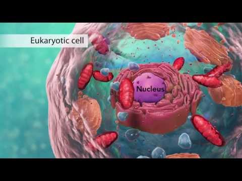
Biology: Cell Structure I Nucleus Medical Media
 0:12:50
Deep learning-based segmentation for biologists - Processing nuclei segmentation - U-Net
 0:09:47
Deep learning for nuclei segmentation and marker identification - Cellpose
 1:11:12
CellProfiler Introduction
 0:28:18
Segmentation of Nucleus and Cytoplasm of a Single Cell in Three-Dimensional Tomogram
 0:01:41
Nuclei segmentation using StarDist in Fiji/ImageJ
 0:00:16
Extreme Cupping Therapy! #shorts #cupping
 0:06:27
Transcription and Translation: From DNA to Protein
 1:00:35
The 3D genome organization and long-range control of gene expression
 0:00:16
NEET 2023 UNEXPECTED RESULT 😞|NEET 2023 SCORE CARD #neet2023 #neet2024 #neetprep #mbbs #short #viral...
 0:03:37
U-net model for nuclei segmentation in medical images
Комментарии