filmov
tv
Introduction to Chest Radiology | Medical Student Online Lectures | V-Learning

Показать описание
Introduction to Chest Radiology explains how to understand the abnormalities of chest through imaging. This V-Learning™ lecture tells about the type of imaging technique and their selection for specific disease, routine chest radiography, and chest radiograph search pattern of variety of diseases.
-------------------------------------------------------------
Lecture Duration: 00:35:21
Released: September 2019
Full List of Radiology Lectures:
-------------------------------------------------------------
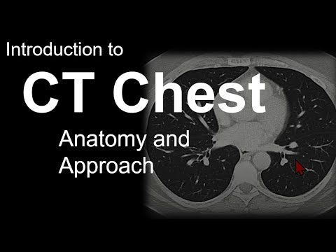
Fundamentals of routine Chest Radiography are discussed in the beginning of the lecture. Then information is given about the imaging techniques which are used to visualize the diseases of the chest. These techniques include chest X-Ray, CT, and MRI along with pulmonary angiography, echocardiography, and nuclear medicine. Likewise, which modality should be used for which kind of disease or symptoms is also explained in succession.
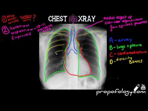
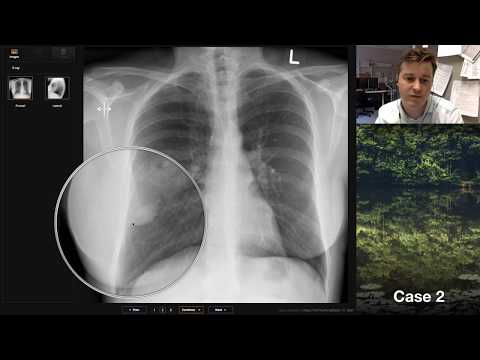
Then representation of PA and lateral chest radiography is given. Lung hila is present in the middle of the lungs whose abnormal pattern is discussed through a radiograph in this lecture later on and is termed as hilar adenopathy. Some Diseases of Pleura such as pleural effusion are also explained through visual representation. Next is the explanation of pulmonary nodules and masses.
Another condition, known as atelectasis, is also described in which decreased expansion of alveoli is observed resulting in collapsed lung. Moving onward, explanation is given on how to interpret the results of Interstitial Lung Diseases on imaging tests. An understandable description is also made on the radiograph demonstration of pectus excavatum. It is a disease of Thoracic Wall in which abnormality in the shape of Sternum and Ribs cage is present.
-------------------------------------------------------------
New medical lectures and clips released every day!
Try for FREE!
-------------------------------------------------------------
Syllabus of Radiology is designed to standardize Radio diagnosis teaching at Post Graduate level resultantly creating competent radiologist with appropriate expertise. For this, Standard books of Radiology are followed i.e. Practical Radiology A Symptom-Based Approach by Edward C. Weber.
-------------------------------------------------------------
Latest Medical Lectures on Facebook:
Latest Medical Lectures on Vimeo:
Latest Medical Lectures on Twitter:
Latest Medical Lectures on Instagram:
Latest Medical Lectures on LinkedIN:
Latest Medical Lectures on TumblR:
Latest Medical Lectures on Pinterest:
-------------------------------------------------------------
-------------------------------------------------------------
Lecture Duration: 00:35:21
Released: September 2019
Full List of Radiology Lectures:
-------------------------------------------------------------
Fundamentals of routine Chest Radiography are discussed in the beginning of the lecture. Then information is given about the imaging techniques which are used to visualize the diseases of the chest. These techniques include chest X-Ray, CT, and MRI along with pulmonary angiography, echocardiography, and nuclear medicine. Likewise, which modality should be used for which kind of disease or symptoms is also explained in succession.
Then representation of PA and lateral chest radiography is given. Lung hila is present in the middle of the lungs whose abnormal pattern is discussed through a radiograph in this lecture later on and is termed as hilar adenopathy. Some Diseases of Pleura such as pleural effusion are also explained through visual representation. Next is the explanation of pulmonary nodules and masses.
Another condition, known as atelectasis, is also described in which decreased expansion of alveoli is observed resulting in collapsed lung. Moving onward, explanation is given on how to interpret the results of Interstitial Lung Diseases on imaging tests. An understandable description is also made on the radiograph demonstration of pectus excavatum. It is a disease of Thoracic Wall in which abnormality in the shape of Sternum and Ribs cage is present.
-------------------------------------------------------------
New medical lectures and clips released every day!
Try for FREE!
-------------------------------------------------------------
Syllabus of Radiology is designed to standardize Radio diagnosis teaching at Post Graduate level resultantly creating competent radiologist with appropriate expertise. For this, Standard books of Radiology are followed i.e. Practical Radiology A Symptom-Based Approach by Edward C. Weber.
-------------------------------------------------------------
Latest Medical Lectures on Facebook:
Latest Medical Lectures on Vimeo:
Latest Medical Lectures on Twitter:
Latest Medical Lectures on Instagram:
Latest Medical Lectures on LinkedIN:
Latest Medical Lectures on TumblR:
Latest Medical Lectures on Pinterest:
-------------------------------------------------------------
 0:07:02
 0:36:54
 0:14:24
 0:24:44
 0:12:37
 0:47:41
 0:11:08
 0:10:30
 0:08:13
 0:16:50
 0:10:11
 0:19:44
 0:07:19
 0:10:30
 0:17:21
 0:05:54
 0:33:01
 0:03:53
 0:11:33
 0:04:34
 0:09:28
 0:41:17
 0:06:11
 0:12:41