filmov
tv
Pharynx Anatomy (Parts, Layers, Muscles)

Показать описание
Content:
0:00 Introduction
0:36 Parts of the Pharynx
1:53 Nasopharynx
7:19 Oropharynx
7:44 Laryngopharynx
8:36 Layers of Pharyngeal Wall
10:17 Muscles of the Pharynx
-------------------------------
-------------------------------
Parts of the Pharynx:
- Nasopharynx, Oropharynx, Laryngopharynx
Layers of the Pharyngeal Wall
Muscles of the Pharynx
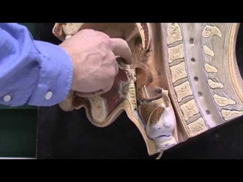
Pharynx:
- 12 to 15 cm long
- Nasopharynx (Pars Nasalis)
- Oropharynx (Pars Oralis)
- Laryngopharynx (Pars Laryngis)
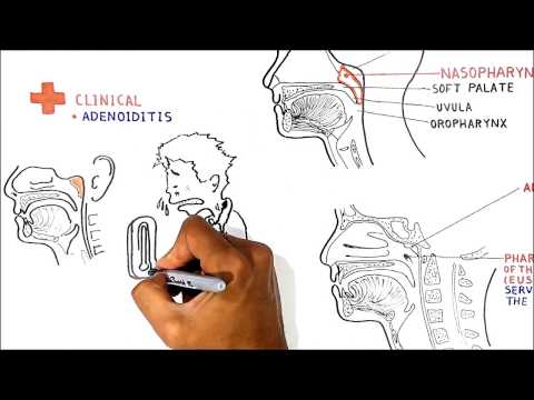
Nasopharynx:
- Level of C1-C2
- Vault of Pharynx (Fornix Pharyngis)
- Attachment points of the pharynx: Pharyngeal Tubercle of the occipital bone (tuberculum pharyngeum), Petrooccipital Fissure (petrooccipital synchondrosis), Inferior Surface of Petrous Part (Temporal Bone), Medial Lamina of Pterygoid Process
- Choana (Internal Nose)
- Auditory Tube
- Pharyngeal opening of the auditory tube (Ostium Pharyngeum Tubae Auditiva)
- Cushion of the Auditory Canal (Torus Tubarius)
- Pharyngeal Recess (Recessus Pharyngeus)
- Pharyngeal Tonsils / Adenoids (Tonsilla pharyngealis)
- Tubal Tonsils (Tonsilla Tubaria)
Overview of Auditory Tube:
- Ear Structures: Outer Ear, Middle Ear (ossicles and tympanic membrane), Inner ear.
- Eustachian Tube connects the nasopharynx with the middle ear
- Function of auditory tube:
○ Equalizing the pressure
○ Draining the middle ear
Oropharynx:
- Level of C4-C4
- Bordered by Soft Palate and Epiglottis
- Connects with the Oral cavity through the Oropharyngeal Isthmus (Isthmus faucium)
Laryngopharynx:
- Level of C5-C6
- Continues into Larynx through Laryngeal Inlet (auditus laryngis)
- Piriform Fossa (Recessus Piriformis)
Layers of the Pharyngeal Wall:
- Tunica Mucosa
○ Lined by Respiratory Epithelium (Pseudostratified epithelium with cilia and goblet cells)
○ Lined by Stratified Squamous non keratinized epithelium
- Tela Submucosa
○ Contains Connective tissue with blood vessels and lymph vessels, and glands
- Tunica Muscularis
○ Has 2 muscle layers for peristalsis
○ Internal Circular muscle layer (Stratum Circulare)
○ Outer Longitudinal Muscle layer (Stratum longitudinale)
- Tunica Adventitia
○ Covers Pharynx from outside
External Pharyngeal Muscles:
- Pharyngeal Constrictors (Musculi Constrictores Pharyngis)
○ Superior Pharyngeal Constrictor
○ Medial Pharyngeal Constrictor
○ Inferior Pharyngeal Constrictor
- Pharyngeal Elevators (Musculi Levatores Pharyngis)
○ Stylopharyngeus Muscle (Musculus Stylopharyngeum)
○ Palatopharyngeus Muscle (Musculus Palatopharyngeum)
○ Salpingopharyngeus Muscle (Musculus Salpingopharyngeus)
Sources used in this video:
- Memorix Anatomy 2nd Edition by Hudák Radovan (Author), Kachlík David (Author), Volný Ondřej (Author)
- Biorender
- University notes and lectures
0:00 Introduction
0:36 Parts of the Pharynx
1:53 Nasopharynx
7:19 Oropharynx
7:44 Laryngopharynx
8:36 Layers of Pharyngeal Wall
10:17 Muscles of the Pharynx
-------------------------------
-------------------------------
Parts of the Pharynx:
- Nasopharynx, Oropharynx, Laryngopharynx
Layers of the Pharyngeal Wall
Muscles of the Pharynx
Pharynx:
- 12 to 15 cm long
- Nasopharynx (Pars Nasalis)
- Oropharynx (Pars Oralis)
- Laryngopharynx (Pars Laryngis)
Nasopharynx:
- Level of C1-C2
- Vault of Pharynx (Fornix Pharyngis)
- Attachment points of the pharynx: Pharyngeal Tubercle of the occipital bone (tuberculum pharyngeum), Petrooccipital Fissure (petrooccipital synchondrosis), Inferior Surface of Petrous Part (Temporal Bone), Medial Lamina of Pterygoid Process
- Choana (Internal Nose)
- Auditory Tube
- Pharyngeal opening of the auditory tube (Ostium Pharyngeum Tubae Auditiva)
- Cushion of the Auditory Canal (Torus Tubarius)
- Pharyngeal Recess (Recessus Pharyngeus)
- Pharyngeal Tonsils / Adenoids (Tonsilla pharyngealis)
- Tubal Tonsils (Tonsilla Tubaria)
Overview of Auditory Tube:
- Ear Structures: Outer Ear, Middle Ear (ossicles and tympanic membrane), Inner ear.
- Eustachian Tube connects the nasopharynx with the middle ear
- Function of auditory tube:
○ Equalizing the pressure
○ Draining the middle ear
Oropharynx:
- Level of C4-C4
- Bordered by Soft Palate and Epiglottis
- Connects with the Oral cavity through the Oropharyngeal Isthmus (Isthmus faucium)
Laryngopharynx:
- Level of C5-C6
- Continues into Larynx through Laryngeal Inlet (auditus laryngis)
- Piriform Fossa (Recessus Piriformis)
Layers of the Pharyngeal Wall:
- Tunica Mucosa
○ Lined by Respiratory Epithelium (Pseudostratified epithelium with cilia and goblet cells)
○ Lined by Stratified Squamous non keratinized epithelium
- Tela Submucosa
○ Contains Connective tissue with blood vessels and lymph vessels, and glands
- Tunica Muscularis
○ Has 2 muscle layers for peristalsis
○ Internal Circular muscle layer (Stratum Circulare)
○ Outer Longitudinal Muscle layer (Stratum longitudinale)
- Tunica Adventitia
○ Covers Pharynx from outside
External Pharyngeal Muscles:
- Pharyngeal Constrictors (Musculi Constrictores Pharyngis)
○ Superior Pharyngeal Constrictor
○ Medial Pharyngeal Constrictor
○ Inferior Pharyngeal Constrictor
- Pharyngeal Elevators (Musculi Levatores Pharyngis)
○ Stylopharyngeus Muscle (Musculus Stylopharyngeum)
○ Palatopharyngeus Muscle (Musculus Palatopharyngeum)
○ Salpingopharyngeus Muscle (Musculus Salpingopharyngeus)
Sources used in this video:
- Memorix Anatomy 2nd Edition by Hudák Radovan (Author), Kachlík David (Author), Volný Ondřej (Author)
- Biorender
- University notes and lectures
 0:13:21
Pharynx Anatomy (Parts, Layers, Muscles)
 0:01:02
The Pharynx Anatomy and Function | Epiglottis + Larynx Examined
 0:03:38
Muscles of the pharynx (preview) - Human Anatomy | Kenhub
 0:19:48
Pharynx I : Internal features
 0:28:30
Structure of Pharynx | Layers | Muscles - Constrictors & Longitudinal | Nerve Supply |Deglutitio...
 0:04:57
Anatomy 4, Mouth, nose, pharynx, swallowing
 0:35:33
Anatomy of the Pharynx, Dr Adel Bondok
 1:07:43
LYMPHATIC SYSTEM & SPECIAL SENSES ANATOMY & PHYSIOLOGY Review
 0:19:52
Pharynx & Larynx - Gross Anatomy
 0:33:43
ANATOMY OF PHARYNX - Location, Extent, Subdivisions, Muscles, Blood and Nerve supply
 0:11:51
Introduction to Anatomy of Pharynx
 0:07:04
Larynx, Pharynx and CST LO 3 - Pharyngeal Muscles
 0:01:38
SWALLOWING OR DEGLUTITION - ANATOMY AND PHYSIOLOGY
 0:15:25
The Pharynx
 0:30:39
Larynx - Membranes, ligaments and muscles - Human Anatomy | Kenhub
 0:01:00
Pharynx in 1 min
 0:18:42
Pharynx Anatomy and physiology |parts, layers, muscles part of digestive and respiratory system|
 0:25:36
Pharynx- Constrictors | Gross Anatomy | NEET PG | Dr. Rohini
 0:48:27
Anatomy of Pharynx
 0:06:17
H&N Part 3 Pharynx
 0:08:05
Pharynx
 0:02:51
Layers of The Pharynx | Wall of The Pharynx | Respiratory System | Pt.13 | Medical Discovery
 0:17:23
Pharynx Anatomy (1/4) | Head & Neck | Anatomy
 0:48:13
ANATOMY OF THE PHARYNX 5 (STRUCTURE)
Комментарии