filmov
tv
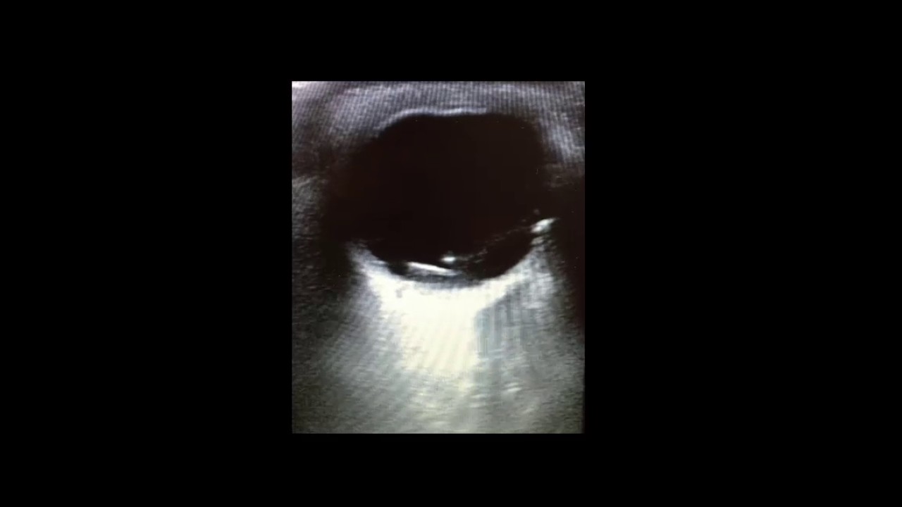
Intraocular Foreign Body, Ultrasound. JETem 2017.
Показать описание
History of present illness: A 36-year-old male with no past medical history presented to the emergency department with left eye pain. Earlier that day, the patient reported pounding a metal object with a metal hammer, when a piece flicked up and struck his eye. He was not wearing eye protection. He noted mild pain. He described his vision as being “dirty water-stained,” seeing “black, floating clouds” and “squiggly lines.” He also described the sensation of something being “stuck” in his eye. On physical exam, his vision was 20/30 in the left eye and 20/20 in the right. His extraocular movements were intact; lid and lash eversion did not reveal a foreign body. There was mild left conjunctival injection, but no tearing or purulent discharge. There was 3 mm of linear corneal fluorescein stain uptake over the anterior chamber.
Significant findings: Point of care ultrasound revealed a mobile, radiolucent hyperechoic structure (see red arrow) with reverberation within the posterior chamber (see blue arrow), likely a metallic foreign body. Linear areas of mobile hyperechoic material revealed possible vitreous hemorrhage (see purple circular area). Orbital non-contrast CT confirmed a 3 mm metallic focus within the dependent portion of the left globe, lodged in the posterior sclera, with some vitreous hemorrhage but no evidence of globe rupture. Ophthalmology was consulted and the patient was taken to surgery later that night.
Discussion: Patients presenting with intraocular foreign body (IOFB) can be easily missed, as they may not complain of vision loss or severe pain. Only a small entry wound may be found on careful examination. However, these seemingly harmless injuries may be vision threatening(1). The IOFB must be removed surgically in the majority of cases (greater than 90%)(2). Bedside ultrasound is a useful tool in looking for metallic objects. Orbital CT continues to be the exam of choice(3).
Topics: Intraocular foreign body, ultrasound and CT of intraocular foreign body, vitreous hemorrhage.
References:
2. Greven CM, Engelbrecht NE, Slusher MM, Nagy SS. Intraocular foreign bodies: management, prognostic factors, and visual outcomes. Ophthalmology 2000; 107:608–612.
 0:00:08
 0:12:40
 0:00:16
 0:29:52
 0:00:18
 0:01:00
 0:00:21
 0:00:44
 0:28:11
 0:00:21
 0:10:24
 0:00:12
![[GH] Lens Dislocation](https://i.ytimg.com/vi/J0hCCIA1tIQ/hqdefault.jpg) 0:01:26
0:01:26
 0:01:18
 0:03:08
 0:00:15
 0:04:28
 0:00:05
 0:00:33
 0:01:19
 0:07:17
 0:04:06
 0:03:58
 0:27:45