filmov
tv
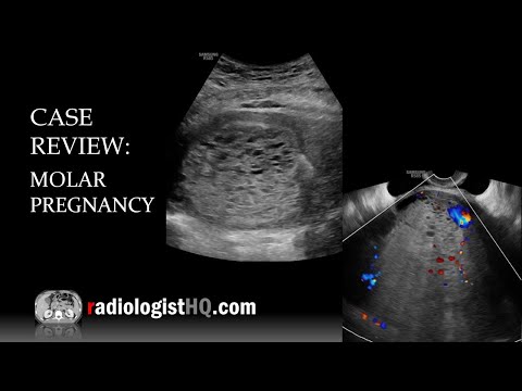
Case Review: Ultrasound of Complete Molar Pregnancy

Показать описание
In this radiology lecture, the ultrasound appearance of complete molar pregnancy is revealed.
Key points include:
1) AKA hydatiform mole = Most common form of gestational trophoblastic disease.
2) Gestational trophoblastic neoplasia (GTN) less common = Invasive mole and choriocarcinoma.
3) Approximately 1/1,000 pregnancies is a molar pregnancy.
4) Most common in females under age 20 and over age 35.
5) Two types of molar pregnancy: Complete (most common) and partial.
6) Complete: Diploid (paternal DNA only), no fetus, more likely to be complicated by GTN.
7) Partial: Triploid (maternal and paternal DNA), abnormal fetus or fetal parts, harder to diagnose.
8) Complete hydatiform mole presentation: Vaginal bleeding, enlarged uterus inconsistent with dates, hyperemesis. Markedly elevated β-hCG level (variable for partial molar pregnancies).
9) Large theca lutein cysts due to ovarian stimulation from elevated β-hCG, but uncommon.
10) US: Heterogeneous, echogenic mass (“snowstorm” appearance), small anechoic cystic spaces (“cluster of grapes”) = hydropic chorionic villi.
11) Treatment: Dilation & curettage. β-hCG levels monitored until no longer detectable to confirm no residual disease.
Click the YouTube Community tab or follow on social media for bonus teaching material posted throughout the week!
 0:05:43
 0:09:57
 0:31:04
 0:08:42
 0:10:22
 0:07:13
 0:05:36
 0:00:06
 1:28:59
 0:05:31
 0:45:23
 0:09:01
 0:06:39
 0:04:24
 0:12:02
 1:11:36
 0:00:10
 0:00:18
 0:20:10
 0:07:11
 0:22:05
 0:31:40
 0:03:47
 0:00:23