filmov
tv
Case of the Week: Perihilar Cholangiocarcinoma/Klatskin Tumor (CT & MRI)

Показать описание
In this radiology lecture, we discuss the CT and MRI appearance of perihilar cholangiocarcinoma.
Key points include:
1) Perihilar cholangiocarcinoma (AKA Klatskin tumor) occurs at bifurcation of the hepatic duct.
2) Cholangiocarcinoma (CC) is a primary malignant tumor of bile duct epithelium, usually adenocarcinoma.
3) CC is the most common primary hepatic malignancy after hepatocellular carcinoma (HCC), and most are extrahepatic (as opposed to intrahepatic).
4) Appearance of CC is based on growth pattern: Mass-forming, periductal infiltrating, and intraductal growing.
5) Risk factors: Parasite infection, choledochal cyst, primary sclerosing cholangitis, recurrent pyogenic cholangitis, and inflammatory bowel disease (ulcerative colitis).
6) Patients are usually 65 or older.
7) On CT and MRI, perihilar CC appears as a biliary stricture with shouldering/abrupt tapering.
8) If a mass is visible, will typically have rimlike enhancement with gradual centripetal enhancement on delayed images, be T2 bright (but not as homogeneous or as bright as hemangioma), and may have a targetlike appearance on DWI (favors CC over HCC)
Click the Community tab or follow on social media for bonus teaching material posted throughout the week!

Case of the Week: Perihilar Cholangiocarcinoma/Klatskin Tumor (CT & MRI)

Case of the Week: Pulmonary Infarction (X-ray & CT)

20 Case of the Week video

Case of the Week: Necrotizing Pancreatitis (CT & MRI)

Case of the Week: Cecal Volvulus (X-ray & CT)

Case of the Week: Large Bowel Lymphoma (CT & PET)

Case-based management of block at the liver hilum and hilar cholangiocarcinoma (Bile duct cancer)

Case of the Week: Retroperitoneal Fibrosis (Ultrasound & CT)

AC21 - Christine Kang: Imaging of Perihilar Cholangiocarcinoma
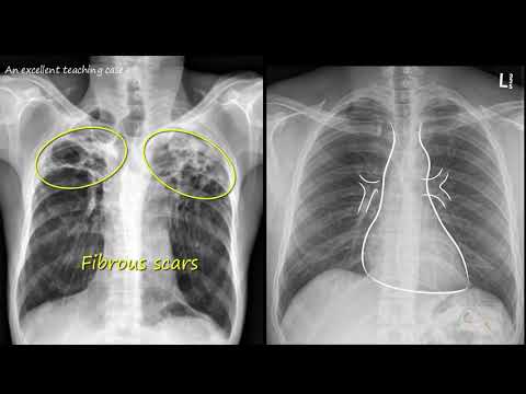
Chest x ray - Tuberculosis healed, (TB), Inactive TB

Inability for detecting cholangiocarcinoma on CT obtained 8 months before diagnosis

AC21 - Ryan Fields: Definition of Perihilar Cholangiocarcinoma

Case of the Week: Medullary Sponge Kidney (Ultrasound & CT)

Case of the Week: Septate Uterus (MRI)

Cholangiocarcinoma | Radiology Everywhere | Radiology Tutorials | #radiology
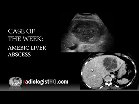
Case of the Week: Amebic Liver Abscess (Ultrasound & CT)

5 Cases in 5 Minutes: Vascular #5

Case 4: 67-Year-Old Woman With Intrahepatic Cholangiocarcinoma

Case of the Week: Wandering Spleen (CT)

Lung Nodules: When to Worry + What to Do Next, Explained by Bronchoscopy Expert Dr. Kyle Hogarth

Interactive CT Abdo Cases - Pancreatitis and Complications (Intro Level)

Case Review: Ultrasound & CT of Renal Oncocytoma

Case of the Week: Gallstone Ileus (X-ray & CT)

Case of the Week: Ovarian Torsion (Ultrasound)
Комментарии