filmov
tv
VERTEBRAL COLUMN ANATOMY (2/2) - Ligaments and the Spinal Cord

Показать описание
Three major ligaments of the spine that allow flexion and extension of the spine while keeping the bones aligned – the ligamentum flavum, anterior longitudinal ligament, and the posterior longitudinal ligament. The anterior and posterior longitudinal ligaments are continuous bands that run from the top to the bottom of the vertebral column and prevent excess movement. The ligament flavum attaches between lamina of each vertebra.
Some additional ligaments I’d like to point out are the intertransverse ligament, the supraspinous ligament, and the interspinous ligament. The intertransverse ligaments stretch between the transverse processes of the spine. The supraspinous ligament is found along the vertebral column, connecting the tips of the spinous processes from the cervical vertebra to the sacrum. At the seventh vertebra, the supraspinous ligament is continuous with the nuchal ligament, which runs from the seventh vertebra to the external occipital protuberance of the skull. The interspinous ligaments are thin, membranous ligaments stretching between adjacent spinous processes.
Finally, I’d like to briefly discuss the spinal cord, which is floating in cerebrospinal fluid within the dural tube, which is inside the vertebral arch. The spinal cord is about the thickness of your thumb. At around 18 inches long, it runs from the brainstem to the 1st or 2nd lumbar vertebra within the spinal canal, ending in the conus medullaris. Extending from the conus medullaris is the cauda equina – Latin for horse tail because it is a bunch of spinal nerves that very much looks like a tail. The cauda equina occupies the lumbar cistern – a space beneath the conus medullaris. The filum terminale extends from the end of the spinal chord and anchors it to the tailbone.
31 pairs of spinal nerves branch off from the spinal cord. Each spinal nerve has 2 roots – one ventral for motor impulses from brain, and one dorsal for sensory impulses to brain. Ventral and dorsal roots fuse to form spinal nerve, which exits the vertebral column via the intervertebral foramen between the vertebrae.
3D model from:
 0:02:11
VERTEBRAL COLUMN ANATOMY (2/2) - Ligaments and the Spinal Cord
 0:04:05
VERTEBRAL COLUMN ANATOMY (1/2)
 0:02:37
Spine Anatomy | Know Your Spine
 0:06:56
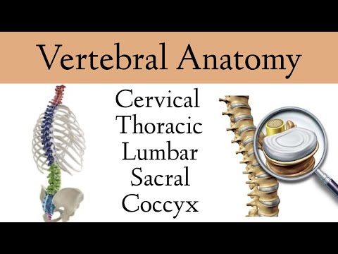
Vertebral Column Anatomy and Bones [Cervical, Thoracic, Lumbar, Sacral Spine]
 0:07:59
Vertebral Column - Introduction | 3D Anatomy Tutorial
 0:09:22
Drawing The SPINE - Anatomy, Structure & Movement - Anatomy 2
 0:09:08
Spine tutorial (2) - Features of a vertebra - Anatomy Tutorial
 0:04:42
Atlas & Axis Cervical Vertebrae (C1-C2) Anatomy
 0:42:37
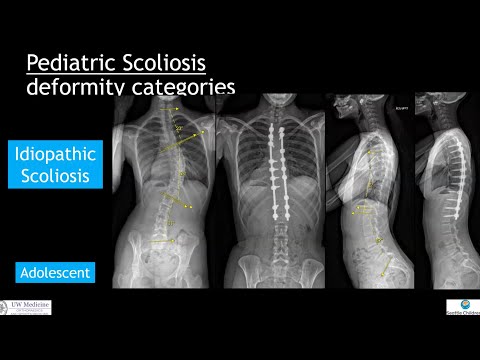
Non-AIS Scoliosis: What's the difference?
 0:00:47
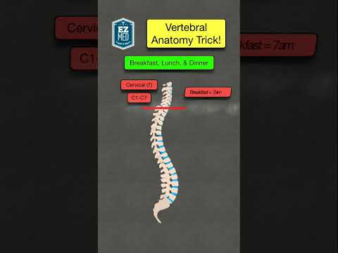
🔥 How to Remember the Anatomy of the Vertebral Column [Spine Diagram]
 0:00:21
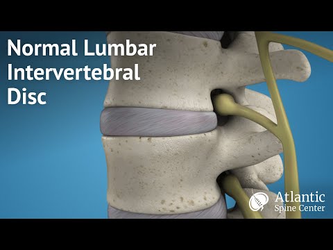
Normal Lumbar Intervertebral Disc
 0:00:43
REAL Human Spinal Cord & Herniated Discs | #shorts #spinalcord
 0:34:22
𝟎𝟒. 𝐕𝐞𝐫𝐭𝐞𝐛𝐫𝐚𝐥 𝐜𝐨𝐥𝐮𝐦𝐧 (𝐏𝐚𝐫𝐭𝐬 𝐚𝐧𝐝 𝐩𝐫𝐢𝐦𝐚𝐫𝐲 𝐚𝐧𝐝 𝐬𝐞𝐜𝐨𝐧𝐝𝐚𝐫𝐲 𝐜𝐮𝐫𝐯𝐞𝐬)...
 0:01:00
Cervical Nerves vs Vertebrae #anatomy #cervicalspine #physiotharapy #physicaltherapy
 0:43:36
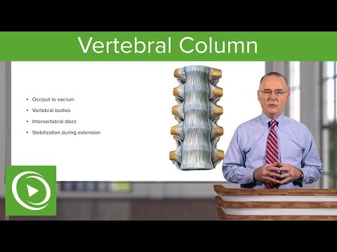
Vertebral Column – Anatomy | Lecturio
 0:06:43
Vertebral Column Anatomy: Bones, Regions, Curvatures (Kyphotic, Lordotic)
 0:43:38
Vertebral Column – Anatomy | Lecturio
 0:00:43
What is Spondylolisthesis? | Vertebral Slippage #Shorts
 0:03:25
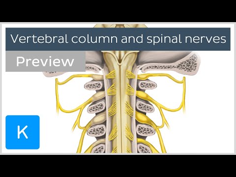
Vertebral column and spinal nerves (preview) - Human Anatomy | Kenhub
 0:18:57
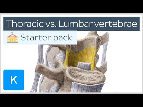
Thoracic vertebrae vs. Lumbar vertebrae - Human Anatomy | Kenhub
 0:03:37
Cervical spine - Anatomy, Diagram & Definition - Human Anatomy | Kenhub
 0:00:59
Spine Anatomy | Know Your Spine #Shorts
 0:00:49
Interesting facts about the human spine!#doctor #anatomy #surgeon #shorts
 0:02:03
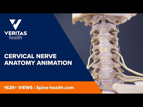
Cervical Nerve Anatomy Animation
Комментарии