filmov
tv
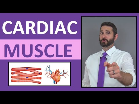
Physiological Anatomy of Cardiac Muscle - Structure & Types

Показать описание
● Follow me at:
Physiological Anatomy of Cardiac Muscle - Structure & Types:
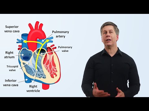
Multiple cardiomyocytes connect at intercalated discs, to make cardiac muscle.
Inside, they have myofibrils, and transverse-tubule system‒ similar to that in skeletal muscle.
The special thing is: the large number of gap junctions at the intercalated discs, make the cardiac muscle a syncytium.
The atrial and ventricular syncytium are separated by fibrous tissue‒ called the atrioventricular ring.
Excitatory and conductive fibers, play a role in controlling rhythm, and don't participate in the contractile process much.
Chapters:
00:00 Intro
00:19 Types of Cardiac Muscle
00:42 Structure of Contractile Muscle Cell
02:08 Intercalated Disc & Syncytium
03:06 Atrioventricular Ring
03:55 Excitatory & Conductive Fibers
04:22 Summary
Dr Vipul Navadiya
Nonstop Neuron
Medical Animation
Medical Animation Videos
Physiology
DISCLAIMER: This video is for education purposes only. Although every effort is made to ensure the accuracy of the material, viewers should refer to the appropriate regulatory body/authorized websites, guidelines, and other suitable sources of information as deemed relevant and applicable. In view of the possibility of human error or changes in medical science, any person or organization involved in the preparation of this work accepts no responsibility for any errors or omissions, or results obtained from the use of information in this video.
 0:05:01
Cardiac Muscle Tissue Anatomy & Physiology Review Lecture
 0:05:20
Physiological Anatomy of Cardiac Muscle - Structure & Types
 0:06:01
Structure of Cardiac Muscle | Cardiac Muscle Tissue | Cardiac Physiology
 0:48:01
Cardiovascular | Electrophysiology | Intrinsic Cardiac Conduction System
 0:10:09
Cardiac Muscle
 0:17:38
Muscle Tissue | Skeletal, Cardiac, and Smooth
 0:07:40
Cardiology - Heart Physiology I (Cardiac Myocyte and Membrane Potential)
 0:06:43
Cardiac Muscle Contraction | Excitation Contraction Coupling | Cardiac Physiology
 0:01:00
Cardiac Muscle Action Potential - Part-1 #shorts #youtubeshorts #youtube #ytshorts
 0:10:08
The Heart, Part 1 - Under Pressure: Crash Course Anatomy & Physiology #25
 0:21:55
Cardiovascular | Structures and Layers of the Heart
 0:07:57
Parts of the Cardiac System (Heart Anatomy)
 0:23:59
Cardiovascular | Cardiac Cycle
 0:28:32
The Cardiovascular System: An Overview
 1:25:36
Ch#09 Physiology Guyton | CARDIAC Muscles | Heart as a PUMP | Cardiac Valves | Cardiac Physiology
 0:10:24
Muscles, Part 1 - Muscle Cells: Crash Course Anatomy & Physiology #21
 0:11:04
Cardiac muscle fiber chap 9 part 1 guyton and hall text book of medical physiology- heart physiology
 0:00:36
Can the Heart Survive Outside the Body?
 0:16:43
Cardiac muscle
 0:48:10
Cardiovascular Physiology - Pressure-Volume loops, Cardiac Cycle, ESV, EDV, SV, CO, Starling Law
 0:06:09
Basic Cardiac Anatomy and Physiology by N. Braudis, A. Olszewski | OPENPediatrics
 0:02:15
Physiology of The Heart: Functional Properties of Cardiac Muscle
 0:08:21
Muscle Tissues and Sliding Filament Model
 0:06:00
Skeletal Muscle Tissue: Contraction, Sarcomere, Myofibril Anatomy Myology
Комментарии