filmov
tv
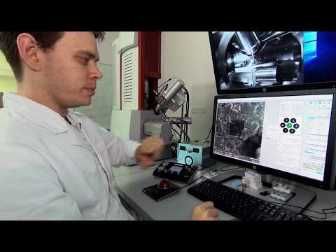
SEM Micrographs Interpretation in Experimental paper: Scanning Electron Microscopy SEM Analysis

Показать описание
How to interpret SEM/FESEM micrographs in your research paper or thesis?
SEM is versatile and a powerful tool for material characterization.
SEM becomes more useful and necessities Due to the continuous shrinking of the material’s dimension for copious applications.
SEM is versatile and a powerful tool for material characterization.
SEM becomes more useful and necessities Due to the continuous shrinking of the material’s dimension for copious applications.
 0:08:13
SEM Micrographs Interpretation in Experimental paper: Scanning Electron Microscopy SEM Analysis
 0:00:53
How Can I Interpret SEM Micrographs in My Experimental Paper?
 0:00:56
How Can I Effectively Interpret SEM Micrographs in My Experimental Paper?
 0:13:27
Scanning Electron Microscope (SEM)
 0:00:58
How Can I Accurately Interpret SEM Micrographs in Experimental Research?
 0:01:01
How Can I Effectively Interpret SEM Micrographs in Experimental Papers?
 0:01:00
How Can I Effectively Interpret SEM Micrographs in Experimental Research?
 0:00:58
How Can I Accurately Interpret SEM Micrographs in Experimental Papers?
 0:00:51
How Can I Accurately Interpret SEM Micrographs in My Experimental Paper?
 0:00:50
How Can I Accurately Interpret SEM Micrographs in My Experimental Paper?
 0:00:53
How Can I Accurately Interpret SEM Micrographs in Experimental Research?
 0:01:05
How Can I Effectively Interpret SEM Micrographs in My Experimental Research?
 0:00:59
How Can I Effectively Interpret SEM Micrographs in My Experimental Paper?
 0:04:37
How to Interpret SEM Images?
 0:00:56
How Can I Accurately Interpret SEM Micrographs in My Experimental Paper?
 0:01:00
How Can I Accurately Interpret SEM Micrographs in My Experimental Research?
 0:00:58
How Can I Effectively Interpret SEM Micrographs in Experimental Research?
 0:00:58
How Can I Accurately Interpret SEM Micrographs in My Experimental Paper?
 0:00:58
How Can I Accurately Interpret SEM Micrographs in My Experimental Paper?
 0:00:48
How Can I Accurately Interpret SEM Micrographs in My Experimental Paper?
 0:00:59
How Can I Accurately Interpret SEM Micrographs in My Experimental Research?
 0:00:52
How Can I Interpret SEM Micrographs in My Experimental Paper?
 0:01:08
How Can I Effectively Interpret SEM Micrographs in an Experimental Paper?
 0:01:02
How Can I Accurately Interpret SEM Micrographs in My Experimental Research?
Комментарии