filmov
tv
Radiology of Nontuberculous Mycobacterium

Показать описание
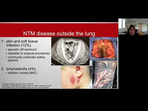
In this video, we talk about the imaging findings of pulmonary nontuberculous mycobacterium (NTM) infection. We discuss both the nodular bronchiectasis form and the fibrocavitary form of the disease with many examples, and discuss a few things to keep in mind in the differential.
Textbooks I like for chest radiology—
Med students and all residents: Felson’s Principles of Chest Roentgenology
Radiology residents: Thoracic Imaging: Pulmonary and Cardiovascular Radiology
Thoracic radiology fellows: Muller’s Imaging of the Chest: Expert Radiology Series
Textbooks I like for chest radiology—
Med students and all residents: Felson’s Principles of Chest Roentgenology
Radiology residents: Thoracic Imaging: Pulmonary and Cardiovascular Radiology
Thoracic radiology fellows: Muller’s Imaging of the Chest: Expert Radiology Series
 0:34:58
Radiology of Nontuberculous Mycobacterium
 0:19:03
Lady Windermere Syndrome (MAC infection) | Radiology Board Review Case
 0:15:37
Board Review | Thoracic Radiology | Part 1
 0:54:46
Nontuberculous Mycobacterial Lung Disease - Dr. Zeid
 0:56:50
Imaging of Pulmonary Tuberculosis
 0:40:45
Treatment of NTM | NTM Lecture Series for Patients and Families
 0:07:36
USMLE DOMINATION: HIGH YIELD TUTORIAL #3- TB
 0:10:43
57 Non tuberculous Mycobacteria Diagnosis&Clinical Management Session 02
 0:44:27
8 Pneumonia TB NTMB
 0:25:34
Jakko van Ingen, MD, PhD - Beyond Skinny Ladies
 0:26:29
Miliary lung pattern - radiology clinical lecture (tuberculosis, fungal lung infections)
 0:53:00
Nontuberculous mycobacterial pulmonary disease and management
 0:27:46
Nontuberculous Mycobacteria NTM - Dr. Ali Dabbagh
 0:23:20
TB and endemic fungi
 0:22:20
Chest - Pulmonary infections the old and the new 1
 0:01:22
Pulmonary MAC infection
 0:03:08
Epituberculosis/Non Tuberculous consolidation of lung - A rare phenomenon (Simplified)
 0:54:56
1/29/21- NTM 2020 Guidelines -- Dr. Hashmi
 1:05:31
Imaging of CNS-Tuberculosis (and other atypical bacterial infections of the brain).
 0:29:04
Overview of Bronchiectasis and NTM
 0:29:25
Radiological evaluation of extra pulmonary TB
 0:02:07
MOOC TUBECULOSIS - Institut Pasteur
 0:55:41
The Changing Face of Nontuberculous Mycobacterial (NTM) Disease: Lady Windermere shares the Stage
 0:13:39
Part 02/03 - Hear the applications of WGS in TB diagnosis from the pride of AIMS | HaystackAnalytics
Комментарии