filmov
tv
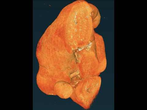
Human Organ Atlas: HiP-CT imaging of a healthy human brain using the ESRF-EBS

Показать описание
Seeing inside a healthy brain using a new imaging technique - Hierarchical Phase-Contrast Tomography or ‘HiP-CT’. Performed at the ESRF-EBS 4th generation synchrotron in Grenoble
HiP-CT is a new technique that can hierarchically image intact whole human organs. Beginning with a scan of the whole organ at the resolution of a human hair (25μm/voxel), followed by zooming in to any area at a resolution of 1/10th a human hair (6μm/voxel), and finally zooming in again to a resolution where we can see single cells (1.5μm/voxel).
The brain is one of the most complex organs, controlling most of our bodily processes. In the video we can initially see the whole human brain, with the two hemispheres at the front and the cerebellum at the back. As we zoom in more features are visible including the grey and white matter and as we zoom in further the small blood vessels become visible and the layers of the cerebellum. The blood vessel network can be seen more clearly in 3D
Results for scientific and medical research and educational use only. HiP-CT and the Human Organ Atlas originated from a group trying to understand how COVID-19 injures our organs. The group are now developing HiP-CT to map our organs in health and disease to better understand them from a whole organ system down to the cellular level.
Project Investigators/contributors: Peter D. Lee and Claire Walsh (UCL), Paul Tafforeau (ESRF), Danny Jonigk (Hannover), Maximilian Ackermann (Mainz), Will Wagner (Heidelberg), Joe Jacob and Simon Walker-Samuel (UCL), Mark Kuehnel and Christopher Werlein (Hannover), Alexandre Bellier (LADAF), and many others helping. We wish to thank ESRF for continuing to support this programme and the development of BM18 led by Paul Tafforeau.
The authors wish to thank the various funders of the authors and this project, including: the Chan Zuckerberg Initiative DAF, an advised fund of Silicon Valley Community Foundation; the Royal Academy of Engineering; the MRC; the Wellcome Trust; the Deutsche Forschungsgemeinschaft; the National Institutes of Health (NIH); and the German Registry of COVID-19 Autopsies (supported by the German Federal Ministry of Health).
 0:02:19
Human Organ Atlas: HiP-CT imaging of a healthy human brain using the ESRF-EBS
 0:01:26
Human Organ Atlas: HiP-CT imaging of a human heart using the ESRF-EBS (2)
 0:00:59
Human Organ Atlas: HiP-CT imaging of a COVID-19 injured human spleen using the ESRF-EBS
 0:01:16
Human Organ Atlas: HiP-CT imaging of a healthy human kidney using the ESRF-EBS
 0:00:45
Human Organ Atlas: HiP-CT imaging of a healthy human lung using the ESRF-EBS
 0:01:37
Human Organ Atlas: HiP-CT imaging of a COVID-19 injured human lung using the ESRF-EBS
 0:02:07
Overview of HiP-CT being used for the Human Organ Atlas using the ESRF-EBS
 0:01:59
Human Organ Atlas: HiP-CT imaging of a Covid-19 injured whole lung lobe using the ESRF-EBS
 0:00:30
Human Organ Atlas: Seeing inside all our major organs at unprecedented resolution using HiP-CT
 0:00:51
Human Organ Atlas Hub (HOAHub) at ESRF Announcement Sept. 2023
 0:52:06
Human Organ Atlas Hub Proposal Town Hall Meeting, Dec. 2, 2022
 0:06:04
Interview with Dr. Claire Walsh on the Human Organ Atlas
 0:04:17
The Human Organ Atlas: a ‘Google Earth’ for our bodies
 0:02:24
HiP-CT at ESRF's BM18, and other BM18 Applications
 0:00:26
Hip CT: Normal (No Pathology)
 0:46:55
BrainMap: Correlative HiP-CT of the human brain to bridge histology and radiology
 0:11:56
PaNOSC presentation by Claire Walsh on the Human Organ Atlas at ESOF 2022
 2:07:56
Presentations from HOAHub virtual workshop May 23rd: How to Analyze HiP-CT Images of Human Organs
 0:08:30
Prof Peter Lee and Dr Claire Walsh: Imaging with HiP CT | MechEng Night 175 - Revolutions
 0:03:25
Super-high-resolution CT visualization
 0:05:23
Pelvis CT anatomy
 1:01:09
24 Hour Human Reference Atlas Event - Hour 21 - Mapping Whole Human Organs at High Resolution
 0:00:21
Hip CT: Lytic Lesions from Multiple Myeloma
 0:11:06
Hip MRI Anatomy Atlas (unlabeled)
Комментарии