filmov
tv
Anatomy and Osteology of Fibula

Показать описание
Mansoor Ahmed
Lower Extremity Anatomy Course Director - Dr. Bareither, PhD
Clinical Anatomy Course Director - Dr. Manion, PhD
Anatomy Lab Diener - Adam Jansen
This video reviews the anatomy and osteology of the fibula. A right bone is used.
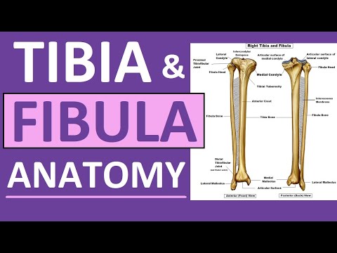
1. The fibula is the lateral bone of the leg. Articulates with tibia forming superior tibiofibular and inferior tibiofibular joint. Long bone that consists of proximal extremity, shaft, and distal extremity.
2. Proximal extremity – also called head of the fibula. Articular facet for the lateral condyle of the tibia. The apex, or styloid process is located posteriorly. This region, where the head meets the shaft, is the neck of the fibula. Proximal fibular fractures can injure the common peroneal (fibular) nerve.
3. Shaft - three borders (anterior, interosseous, posterior) and three surfaces (medial, lateral, posterior). Nutrient foramen and crista medialis. FHL originates lateral to the crista medialis, and the tibialis posterior takes part of its origin medial to the crista.
4. Distal extremity - inferior to medial malleolus. It is composed of two surfaces, two borders, and an apex.
5. Medial surface - triangular shaped, base up/apex down articular facet for talus. The posterior talofibular and inferior transverse ligaments attach to the malleolar fossa.
6. Anterior Border - anterior talofibular and calcaneofibular ligaments
7. Apex – calcaneofibular ligament
8. Posterior Border - lateral malleolar sulcus, which allows passage for the tendons of peroneus longus and brevis.
9. Primary Center of Ossification – 6th to 7th month of fetal development in shaft
10. Secondary Center of Ossification – 4th year of age in proximal extremity. 1 to 1.5 years of age in distal extremity
Lower Extremity Anatomy Course Director - Dr. Bareither, PhD
Clinical Anatomy Course Director - Dr. Manion, PhD
Anatomy Lab Diener - Adam Jansen
This video reviews the anatomy and osteology of the fibula. A right bone is used.
1. The fibula is the lateral bone of the leg. Articulates with tibia forming superior tibiofibular and inferior tibiofibular joint. Long bone that consists of proximal extremity, shaft, and distal extremity.
2. Proximal extremity – also called head of the fibula. Articular facet for the lateral condyle of the tibia. The apex, or styloid process is located posteriorly. This region, where the head meets the shaft, is the neck of the fibula. Proximal fibular fractures can injure the common peroneal (fibular) nerve.
3. Shaft - three borders (anterior, interosseous, posterior) and three surfaces (medial, lateral, posterior). Nutrient foramen and crista medialis. FHL originates lateral to the crista medialis, and the tibialis posterior takes part of its origin medial to the crista.
4. Distal extremity - inferior to medial malleolus. It is composed of two surfaces, two borders, and an apex.
5. Medial surface - triangular shaped, base up/apex down articular facet for talus. The posterior talofibular and inferior transverse ligaments attach to the malleolar fossa.
6. Anterior Border - anterior talofibular and calcaneofibular ligaments
7. Apex – calcaneofibular ligament
8. Posterior Border - lateral malleolar sulcus, which allows passage for the tendons of peroneus longus and brevis.
9. Primary Center of Ossification – 6th to 7th month of fetal development in shaft
10. Secondary Center of Ossification – 4th year of age in proximal extremity. 1 to 1.5 years of age in distal extremity
 0:03:40
Anatomy and Osteology of Fibula
 0:08:32
FIBULA | BONES OF LOWER LIMB | ANATOMY | SIMPLIFIED ✔
 0:05:47
Tibia and Fibula Anatomy of Leg Bones | Anatomy & Physiology
 0:05:26
Tibia and Fibula
 0:11:27
Osteology of Fibula
 0:11:36
Tibia and Fibula: Skeletal Anatomy
 0:12:01
Fibula
 0:03:32
FIBULA - SIDE DETERMINATION
 0:16:30
FIBULA - GENERAL FEATURES AND ATTACHMENTS
 0:02:11
Osteology of the Fibula – Anatomy | Lecturio
 0:05:24
Fibula Bone - Introduction, Anatomy, Function, Injuries and Treatment.
 0:09:26
fibula bone anatomy 3d | anatomy of fibula bone attachments anatomy | bones of lower limb anatomy
 0:03:21
Tibia and fibula (preview) - Human Anatomy | Kenhub
 0:36:22
Chp2 | Tibia & Fibula| Tibia | Fibula | Lower Limb | BD Chaurasia Anatomy | Dr Asif Lectures
 0:24:53
Osteology of the tibia, fibula, And Foot (In HD)
 0:12:45
Osteology of Fibula
 0:08:59
Fibula - Osteology
 0:15:24
Osteology of Fibula
 0:00:41
Visual Anatomy 3D: Fibula Facts
 0:17:31
The Tibia
 0:06:29
Fibula Bone 3D anatomy model animation, Side determination, relation with tibia bone , Osteology
 0:00:10
Tibia Fibula Bones
 0:13:42
Osseous Anatomy of the Fibula (Advanced)
 0:13:34
Osteology of Fibula and its attachments
Комментарии