filmov
tv
Sinonasal Anatomy
Показать описание
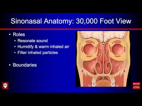
In this video, I discuss Sinonasal Anatomy from the perspective of a neuroradiologist focusing on the CT imaging appearance of the nose, nasal cavity, paranasal sinuses, and frontal recess cells. I also detail the normal sinonasal outflow pathways and important anatomic variants, before concluding with a review of important imaging findings that should be evaluated and reported prior to endoscopic sinus surgery. I think this tutorial will be of value to radiology learners and hope you enjoy it!
All educational content herein has been fully de-identified to protect patient privacy.
All educational content herein has been fully de-identified to protect patient privacy.
Sinonasal Anatomy
 0:07:55
Clinical Anatomy - Nasal Cavity and Sinuses
 1:11:30
Imaging Anatomy of the Paranasal Sinuses
 0:11:34
Paranasal Sinuses and Nasal Cavity | Radiology anatomy part 1 prep | CT imaging
 0:11:01
Nasal Anatomy (Cartilage, Nasal Cavity, Sinuses, Meatuses, Nasal Mucosa)
 0:58:21
Rhinology | Radiology of the nose and paranasal sinuses | Dr Steve Connor
 0:09:36
Basic CT Anatomy of Paranasal Sinuses, Made Easy
 0:47:22
Rhinology | Sinonasal Radiology | Dr Ashok Adams
 0:36:05
‘GRN-ACADEMY' - ESS, Module 1 - Topic 2 - Clinically relevant Sinonasal Anatomy - Dr Timothy Be...
 0:06:02
Chapter 1: Basic Sinus Anatomy
 0:03:13
ANATOMICAL VARIATIONS IN SINONASAL REGION (CT SCAN OVERVIEW)
 0:43:36
2-Anatomy of sinonasal unit
 0:01:55
Sinusitis Surgery
 0:10:45
How to read a Sinus CT
 0:56:08
3-Pathology of the sinonasal unit I
 0:33:03
CT Nose and Paranasal Sinuses Anatomical View Part 1
 0:23:16
Pre-op FESS Checklist - Dr. Suresh Mukherji - Medality (MRI Online) Radiology Noon Conference
 0:02:17
Anatomy of maxillary sinus
 0:30:36
01. Imaging Anatomy of Paranasal Sinuses
 0:30:50
045. Anatomy Maxillary Sinus. #anatomylectures
 0:06:28
Sinusitis - causes, symptoms, diagnosis, treatment, pathology
 0:02:12
Anatomy of the Paranasal Sinuses
 0:55:35
Anatomy of Nose and Paranasal Sinuses
 0:26:56
Imaging of the Paranasal Sinuses 3
Комментарии