filmov
tv
Virtual Journey Through the Heart

Показать описание
Fraunhofer MEVIS - Institute for Digital Medicine
Public since August 7, 2015
2D preview of 3D movie in the Deep Space
David Black, Anja Hennemuth, Bianka Hofmann, Alexander Köhn, Mathias Neugebauer, in cooperation with Andreas H. Mahnken, University Hospital Gießen and Marburg, Germany
On August 7, 2015 the brand new Deep Space in the Ars Electronica Center in Linz, Austria will reopen its gates. The Deep Space is a 16 x 9 meter large projection canvas and has been upgraded with high-end 8K Stereo Shutter technology. Fraunhofer MEVIS has contributed a movie to the new programm 'Universum Mensch'.
Deep Space
Description:
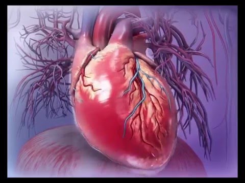
CT image layers build up a three-dimensional block. The bright pixels represent dense structures that have absorbed many X-rays and therefore appear white in the CT images. Colored medical image data visualizes anatomical structures. A rhythmically beating human heart can be seen. The sternum, ribs, and diaphragm underneath the heart are colored turquoise. The right heart muscle pumps the blood gathered from the body through the blood vessels into the lungs. The left heart muscle draws the oxygen-enriched blood from the lungs and pumps it into the entire body. The sinoatrial node determines the heart beat’s rhythm. It starts the electrical stimulation of the healthy person’s heart muscle cells. This stimulation spreads downward.
0:58
Now back to the two-dimensional view of a CT layer, which doctors use to determine whether the heart valves, depicted in blue, function properly. The distinctively pumping left heart muscle distributes the oxygen-enriched blood from the lungs into the entire body. The heart valves absorb less radiation energy and therefore appear dark in the CT image. Muscles and tissues receive, in contrast, more radiation and appear in different shades of gray.
An elevation map emerges based on the pixels’ gray values. The brighter the area, the higher the hill, the more radiation a pixel has received.
1:42
Instead of using (showing) the intensity of the radiation in CT images, brightness can encode the blood flow velocity in MR images. Scientists have found a way to graphically present complex information about motion gained from MR pixels. Blood flows the fastest in the orange areas. Similar to ink spreading in water, virtual particles visualize the direction of the blood flow. Their movement reconstructs the blood flow in the patient’s heart step by step. The heart muscle can be clearly seen in anatomical MR images.
In the future, these new imaging methods could help doctors determine how the blood flow changes due to heart diseases, without using a catheter. They help calculate how the blood pressure and shear forces on the wall of the blood vessels have changed for patients with heart valve problems. In the future, mathematical simulations will help estimate the benefits provided by a new heart valve.
The three-minute sequence is based exclusively on real medical data gathered by the Institute’s MRI scanner. The CT data were provided by the University Clinic in Marburg. Fraunhofer scientists created the movie using “MeVisLab”, a software package for developing medical image processing assistance systems for physicians. Experts and industrial partners around the world currently use MeVisLab.
 0:02:54
Virtual Journey Through the Heart
 0:02:54
Virtual Journey Through the Heart (Voice-Over)
 0:00:45
Travel Through the Human Heart Using Virtual Reality
 0:06:24
A Journey Through the Heart
 0:04:00
Swim Along the Arteries and Through the Heart as part of the Blood Stream in 360° VR
 0:03:10
The Path of Blood Through the Heart
 0:04:24
Visible Body | Virtual 3D Human Heart Anatomy Walkthrough
 0:08:07
WHAT HAPPENS INSIDE YOUR BODY? || 360 VR
 0:26:08
OSLO NORWAY, Autumn Walk In Big City Oslo🇳🇴 Virtual Walk Tour’ 4K/60ftp
 0:01:06
VR 360 Animation - Inside the Human Body
 0:15:19
Your Personal Virtual Heart | Natalia Trayanova | TEDxJHU
 0:10:27
Journey to the Center of the Milky Way Galaxy Like Never Before (4K)
 0:02:46
Human Digestive System in VR!!! | Education in 360
 0:42:45
Journey to the Edge of the Universe [4K]
 0:03:31
Three minutes to the centre of the Earth - BBC
 0:06:10
Virtual Nature 360° - Nature Meditation for VR Quest
 0:15:00
🌎 SIMULATED Journey from EARTH to the END of the UNIVERSE ✨
 0:03:40
Escape Now: Florence & Rome in 360° VR | A Guided Journey Through Italy's Artistic Heart
 0:08:22
What Happens Inside Your Eyes - 3D Animation
 0:04:32
Escape Now: Paris in 360° VR | An Enchanting Guided Journey Through the City of Lights
 0:03:52
360 / VR Video - Experience Going to Heaven / The Afterlife
 0:03:07
Escape Now: London in 360° VR | A Timeless Guided Tour Through England's Historic Capital
 1:02:20
A Relaxing Virtual Walk Through the Central Streets of the Most Enchanting City in the World.
 0:02:37
Center for Healthcare Education Virtual Tour | Nursing
Комментарии