filmov
tv
Radiation causing cancer (radiation carcinogensis for Radiologic Technologists)

Показать описание
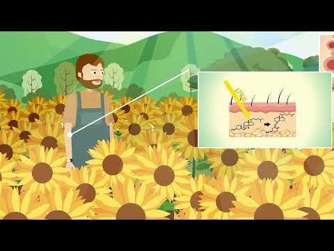
Radiation carcinogenesis (a fancy way of saying a higher risk of cancer induction after being exposed to ionizing radiation). The mechanism for carcinogenesis is primarily x-rays coming in, generating energetic electrons which then either directly or indirectly cause DNA damage. The data which we have on carcinogenesis is from high radiation dose events such as the atomic bomb survivors and there is not complete consensus on the best way to estimate the effects of the very low levels of radiation, which are received during diagnostic procedures. The most accepted method being the linear no threshold model which directly fits a line to the data that is available and uses this to extrapolate what the effects will be for low levels of radiation.
Chapters:
00:00 Intro
00:58 Radiation Trailblazers
01:55 Atomic bomb survivors
03:02 Cancer examples due to radiation
05:30 Extrapolated Risks of Radiography
06:28 Linear No Threshold (LNT)
When the DNA cannot be properly repaired there is some chance of cancer induction. Here we discuss the cases in human history where a clear link has been demonstrated between radiation dose and cancer risk.
All of the solid data on elevated cancer risk is from relatively high dose exposures. Therefore, in order to estimate the risk at the low radiation doses of diagnostic x-ray and CT scanning a method is needed to estimate data beyond the measurements.
The process of estimating data beyond the available measurements is called extrapolation, and multiple methods for performing the extrapolation are presented.
Marie Curie, famous physicist/chemist and two-time Nobel Prize laureate discovered fundamental properties involving radioactive isotopes. She is believed to have died from leukemia.
Additionally, early doctors, dentists and x-ray technologists who had been working with radiation before the downsides and the health effects of radiation were known also had significantly higher risks of cancer than the normal population.
Atomic Bomb Survivors (primary knowledge of radiation/cancer link)
Most information about radiation and its effects on humans comes from atomic bomb survivors. After the two explosions of atomic bombs in Hiroshima and Nagasaki Japan in 1945.
Leukemia, blood cancer, has been developed in atomic bomb survivors. Another example is liver cancer that has been demonstrated to have a higher prevalence in atomic bomb survivors.
Atomic bomb survivors from Hiroshima and Nagasaki also have seen increased risk of thyroid cancer. And the Chernobyl accident also demonstrated increased risk of thyroid cancer.
For the purpose of estimating the cancer induction risk due to diagnostic radiology procedures we need to determine what the impact is in the far bottom left-hand corner of this type of graph. These exams are typically on the order of several milliSieverts (mSv).
There is not actual good data in this area of the graph so we have to do something called extrapolation to estimate the risk of cancer induction due to these diagnostic procedures.
The simplest technique is to draw a straight line through the data. The blue line here is what we get if we draw a straight line through the data and then we keep drawing that straight line down all the way to the origin where there’s zero radiation dose.
That line is called linear extrapolation and in this case we also say there’s no threshold. So, there’s no threshold below which the effects change. Even at a very low level, we assume the effects are the same. For this reason the most commonly accepted extrapolation method is referred to as Linear No Threshold (LNT).
The final option is called hormesis. In that case, the extrapolated curve actually goes below the x-axis indicating that at very low levels, there’s actually a positive effect of the radiation dose.
High Dose, High Dose Rate Low Dose, Low Dose Rate
Working Population 0.08 / Sv 0.04 / Sv
Entire Population 0.01 / Sv 0.05 / Sv
ICRP Summary value of elevated risk of dying of cancer due to radiation exposure.
To summarize, this currently accepted approach in the field is to use the simplest approach of drawing the straight line through the data and use a linear extrapolation.
Using this approach the ICRP guidelines are given in this figure. In a working population, if there is a relatively high radiation dose, it’s about an 8% increase in cancer induction per Sv. Keep in mind that the doses for diagnostic exams are typically reported in mSv (1/1000 of a Sv).
For a relatively lower radiation dose, like a diagnostic procedure, it’s about a 4% increase and this is per Sievert.
 0:10:04
Radiation causing cancer (radiation carcinogensis for Radiologic Technologists)
 0:10:38
Radiation Carcinogenesis
 0:05:21
Is radiation dangerous? - Matt Anticole
 0:04:46
How UV Causes Cancer and Aging
 0:05:11
How Does Radiation Cause Cancer? | What if you're Exposed to the Highest Doses of Radiation?
 0:35:04
Radiation Carcinogenesis
 0:04:14
Cancer Risk From CT Scan Radiation
 0:35:04
M-22. Radiation Carcinogenesis
 0:01:00
Radiation Effects Are Underestimated
 0:39:08
Lecture: radiation induced cancers
 0:22:20
Radiation and Cancer -by Richard Steeves, MD, PhD @ TEAC7
 0:20:58
✅✅CHEMICAL AND RADIATION CARCINOGENESIS II CHAPTER 7 II NEOPLASIA II ROBBIN 10TH E II PATHO LECTURE...
 0:04:56
Late and long-lasting effects of cancer treatment
 0:10:11
Do CT scans (computed tomography scans) cause cancer?
 0:28:00
Neoplasia:Chemical, Radiation, Microbial carcinogenesis, Screening Methods
 0:20:35
Study to Estimate Radiation Doses and Cancer Risks from Exposure to Fallout from the Trinity Test
 0:00:54
Dr Ljubica Zupunski on cancer and ionizing radiation
 0:02:13
How do radio frequency radiation and electromagnetic fields affect human beings?
 0:03:50
Radiation induced coronary artery disease – RICAD
 0:02:58
Radiation Carcinogenesis; Ultraviolet rays and Ionizing Radiation
 0:00:20
Can📱 radiation cause breast cancer?
 0:01:12
Radiation carcinogenesis
 0:24:20
David J. Brenner, Estimating Cancer Risks at Very Low Radiation Doses
 0:00:11
Macrophage eating Cancer Cell 3D Animation | Phagocytosis | Immunology @biologyexams4u
Комментарии