filmov
tv
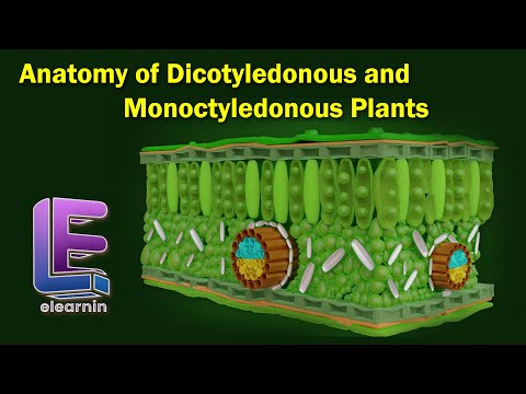
Anatomy of Monocot and Dicot stems

Показать описание
Anatomy of monocot and dicot stems:
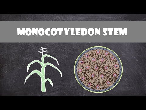
Primary structure of monocot stem - Maize stem:
The outline of the maize stem in transverse section is more or less circular. Internal structure of monocotyledonous stem reveals epidermis, hypodermis, ground tissue and vascular bundles.
Epidermis:
It is the outermost layer of the stem. It is made up of single layer of tightly packed parenchymatous cells. Their outer walls are covered with thick cuticle. The continuity of this layer may be broken here and there by the presence of a few stomata. There are no epidermal outgrowths.
Hypodermis:
A few layer of sclerenchymatous cells lying below the epidermis constitute the hypodermis.. This layer gives mechanical strength to the plant. It is interrupted here and there by chlorenchyma cells.
Ground tissue:
There is no distinction into cortex, endodermis, pericycle and pith. The entire mass of parenchymatous cells lying inner to the hypodermis forms the ground tissue. The cell wall is made up of cellulose. The cells contain reserve food material like starch. The cells of the ground tissue next to the hypodermis are smaller in size, polygonal in shape and compactly arranged. Towards the centre, the cells are loosely arranged, rounded in
shape and bigger in size. The vascular bundles lie embedded in this tissue. The ground tissue stores food and performs gaseous exchange.
Vascular bundles:
Vascular bundles are scattered in the parenchymatous ground tissue. Each vascular bundle is surrounded by a sheath of sclerenchymatous fibres called bundle sheath. The vascular bundles are conjoint, collateral,
endarch and closed. Vascular bundles are numerous, small and closely
arranged in the peripheral portion. Towards the centre, the bundles are
comparatively large in size and loosely arranged. Vascular bundles are skull shaped.
Phloem:
The phloem in the monocot stem consists of sieve tubes and companion
cells. Phloem parenchyma and phloem fibres are absent. It can be distinguished into an outer crushed protophloem and an inner metaphloem.
Xylem:
Xylem vessels are arranged in the form of the letter ‘Y’. The two metaxylem vessels are located at the upper two arms and one or two protoxylem vessels at the base. In a mature bundle, the lowest protoxylem disintegrates and forms a cavity known as protoxylem lacuna.
Disclaimer
This channel does not promote or encourage any illegal activities.
All contents provided by this channel for general and education purpose only.
Copyright disclaimer under section 107 of the copyright act 1976,allowance is made for "fair use policy" for purposes such as criticism, comment, news reporting,teaching,scholarship and research. Fair use is a use permitted by copyright statute that might otherwise be infringing. Non-profit, educational or personal use tips the balance in favor of fair use.
 0:05:53
Anatomy of Dicotyledonous and Monoctyledonous | Anatomy of Flowering Plants | Class 11 Biology
 0:03:44
MONOCOT vs DICOT | Differences between Monocotyledon and Dicotyledon with Examples | Science Lesson
 0:04:20
Anatomy of Monocot and Dicot stems
 0:05:59
Characteristics of Dicot and Monocot Stem and Root - MeitY OLabs
 0:09:05
Ch-6 Anatomy of Root | Dicot root Vs Monocot root | Class 11 Biology/NEET/AIIMS
 0:01:14
MONOCOT & DICOT ROOT COMPARISONS
 0:46:40
CBSE Class 11 Biology || Anatomy of Flowering Plants || Full Chapter || By Shiksha House
 0:09:33
Ch-6 Anatomy of Stem | Dicot Vs Monocot stem | Class 11 Biology /NEET/AIIMS
 0:00:46
Fundamental defferens bw #monocot #Dicot #seeds #biology
 0:01:29
MONOCOT & DICOT STEMS COMPARED
 0:02:17
Monocot Stem Structure and Function | Plant Biology
 0:03:39
Monocot Seed anatomy, Structure of Monocot seed, Monocot Seed, Biology
 0:14:31
Anatomy of Root (Dicot & Monocot) | Anatomy of Flowering Plants| NEETPremier | AA Ma'am | E...
 0:17:30
Week 8 Anatomy of Dicot and Monocot Roots
 0:01:49
MONOCOT & DICOT LEAVES COMPARED
 0:02:41
Anatomy of a Dicot and Monocot Leaves
 0:09:36
Ch-6 | Anatomy of Leaf | Monocot Vs Dicot Leaf | Anotomy of Flowering Plants | Class 11 Biology/NEET
 0:04:00
Dicot stem | sunflower stem practical | Dicot stem anatomy
 0:02:20
Anatomical differences between dicot stem and monocot stem
 0:56:23
Anatomy of Dicotyledonous & Monocotyledonous Plants | Class 11 NEET Biology | NEET 2020 | Vedant...
 0:21:21
Plant Anatomy 6 | Leaf Anatomy | Dorsiventral vs Isobilateral Leaf | Dicot vs Monocot Leaf
 0:03:48
Monocot Root Anatomy, | Maize root , Root Tissue | Journey Inside Maize Roots 11th Bio Practical 🔬🌽&...
 0:10:13
T.S. of Dicot and Monocot root | Plant Anatomy | V Senthilnathan
 0:15:15
Ch-6 -Anatomy of Flowering Plants| Secondary Growth |Vascular & Cork Cambium |Class 11 Biology/N...
Комментарии