filmov
tv
Live Human Blood Cells (erythrocytes and leukocytes). A Neutrophil eating bacteria. DIC Microscopy

Показать описание
This video showing blood cells under the microscope i.e. red (erythrocytes) and white (neutrophils) cells. It also shows how a neutrophil "eats" bacteria (micrococci) - a process called phagocytosis.
Two setups of differential interference contrast (DIC) are tested: the old one with a cheap analyzer from ebay + a zeiss aplanatic/achromatic condenser (NA: 0.9), and the new one with original zeiss analyser + zeiss aplanatic/achromatic condenser (NA: 1.4 oil). I think the difference between the two setups is obvious. What do you think?
I do not mean to cover up lack of microscopy skills, but the image is much clearer when I look through the eyepiece, probably due to the camera...my next investment project. Any suggestions?
Microscope: Axioplan 1.
Objectives: Mostly Zeiss 63x/1.4 Plan-apochromat and Zeiss 100x/1.3 Plan-neofluar in the end.
#microscopy, #/WBC, #BloodCells,
#hematology
 0:04:35
Live Human Blood Cells (erythrocytes and leukocytes). A Neutrophil eating bacteria. DIC Microscopy
 0:06:14
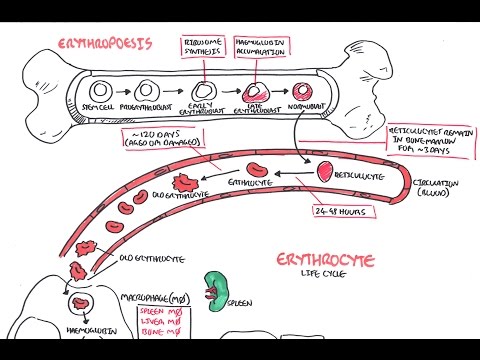
Haematology - Red Blood Cell Life Cycle
 0:03:48
Red blood cells under the microscope, hypo and hypertonic solutions
 0:00:53
White blood cells fight infection under the microscope! RARE FOOTAGE!
 0:04:51
Understanding Erythropoiesis
 0:01:00
Ridiculous Facts About Red Blood Cells!
 0:00:59
White Blood Cell Fights GIANT GERM!
 0:00:28
Human Blood Under Microscope! Watch All the Blood Cells ! #biology #mbbs #neet
 0:05:58
Live Human Blood Cells - RBC, White Blood Cells/Leukocytes, and Platelets. DIC Microscopy (4K)
 0:02:08
Red Blood Cells under the Microscope 400X and 1000X - Part 1
 0:00:16
human blood under the microscope #laboratory #medtechstudent #mls #cls #bloodcells #microscope #bld
 0:00:58
White Blood Cells fighting under microscope!
 0:01:19
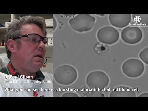
Malaria parasites invading human red blood cell
 0:00:16
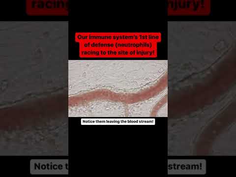
White blood cells leaving the bloodstream (Extravasation)
 0:00:56
A white blood cell's final battle under a microscope!
 0:00:47
Blood vs Bacteria microscope! Insane!
 0:12:00
Parasites in Live Blood Cell
 0:03:18
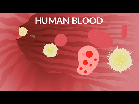
Human Blood Video | Blood Components | Blood Cells
 0:00:16
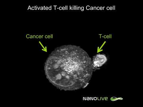
Label-free Live Cell Imaging: Activated T-Cell Killing Cancer Cell
 0:00:29
Real blood under microscope is insane!
 0:00:29
White Blood Cells: Our Body's Silent Killers
 0:00:14
live human blood cell #redbloodcells #shorts #microscope
 0:10:08
Identifying Leukocytes | Review and Practice
 0:00:28
Label-free Live Cell Imaging: T-cells killing cancer cells - zoomed-in
Комментарии