filmov
tv
What blood looks like down the microscope

Показать описание
Can you tell the difference between a monocyte and a basophil? Would you know what an eosinophil looked like if it came up to you and introduced itself? Where do platelets come from? If I have worms, what cells are responsible for killing them?
All this and much more answered as Susan Anderson takes you on a tour of blood on a microscopic scale, teaching you how to identify red cells, granulocytes, lymphocytes, platelets, monocytes, basophils, eosinophils and neutrophil polymorphs and going through what each cell type is for.
Don't miss this if you are a medical student or doctor taking early postgrad exams.
Susan Anderson is Associate Professor of Surgery at the University of Nottingham, UK
 0:07:04
What blood looks like down the microscope
 0:00:58
White Blood Cells fighting under microscope!
 0:00:51
Red blood cells #shorts
 0:00:15
What to Do If You See Blood in Your Stool #jeffersonhealth #gastroenterology
 0:00:26
How Are Blood Clots Removed
 0:03:17
Blood in your poop: what it looks like & what it could mean
 0:00:29
Try this simple blood circulation test #shorts
 0:00:17
Forming of a Blood clot #science #medical
 0:00:11
Should You Pop This Blood Blister? #shorts #blister
 0:00:20
Did You Wake Up With Blood In Your Eye? | Eye Doctor Explains #shorts #health #eyes
 0:05:43
7 Warning Signs of a BLOOD CLOT (Symptoms) 2024
 0:00:51
White blood cell attacks a yeast cell!
 0:00:22
#shark #attack #shorts #ocean #blowthisup #blood #scary #viral #trending #fyp #foryou #popoff
 0:02:12
Warning signs of blood clotting
 0:00:42
Do you think you have a blood clot?
 0:00:36
BLOOD in URINE?! What should YOU do!
 0:00:42
The Sky In China Just Turned Blood Red
 0:05:29
Which Blood Pressure Reading is More Important, Systolic or Diastolic?
 0:09:44
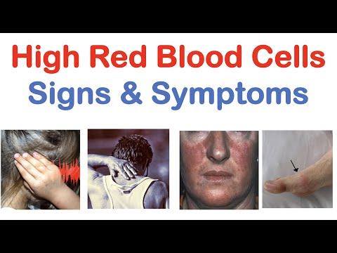
High Red Blood Cells (Polycythemia) Signs & Symptoms (& Why They Occur)
 0:00:27
period blood flow😱😲😱🩸🩸/ #periods #bloodflow/ blood flow during periods/#menstruation
 0:01:09
Mayo Clinic Minute: Breaking down different types of blood donations
 0:06:59
Period Blood Colors Explained | Myths | What It Says About Your Health
 0:02:03
Blood Plasma Problems
 0:00:58
The Top 3 Blood Test Markers To Get For Men Concerned About Testosterone
Комментарии