filmov
tv
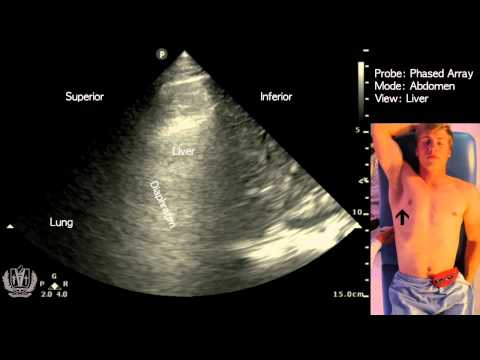
Liver Ultrasound Protocol

Показать описание
Liver Protocol basics.
The liver is the largest organ in the body, the right lobe is 5-6 times larger than the left.
Normally measures about 15cm at mid clavicular line in adults, there is much variation in pediatric populations by age and charts do exist.
The liver is covered by a layer of connective tissue referred to as Glisson's capsule. The liver is normally more echogenic then the renal cortex.The liver is drained by the hepatic veins of which there is usually 3, Left, Middle and Right. They connect to the IVC which drain into the right atrium.
The liver is fed oxygenated blood by the hepatic artery and portal vein.
Excellent article detailing Couinad's segmental liver anatomy
The Radiology Assistant : Anatomy of the liver segments
 0:07:26
Liver Ultrasound Protocol
 0:08:11
Liver Ultrasound Probe Positioning | Transducer Placement For Liver Scanning | Abdominal USG
 0:09:57
Liver Ultrasound Scanning Protocol | AIMS Education
 0:05:36
How to ultrasound the liver
 0:23:40
Liver Protocol :: Sononerds:: Doodle Tutor
 0:02:45
Transverse Liver Scan Technique
 0:02:01
Liver Ultrasound Protocol In Transverse Scan
 0:04:34
Point of Care Ultrasound of the Gallbladder - AMBOSS Video
 0:10:00
Basic Sonographic Anatomy of the Liver
 0:12:02
US Abdomen Complete Protocol
 0:03:03
Abdominal Ultrasound - Basics of Evaluating the Liver
 0:02:45
How To Measure Liver On Ultrasound | Craniocaudal Length, Transverse, Volume & AP Measurements U...
 2:05:07
Liver: Anatomy, Physiology & Normal Ultrasound :: Unit 3 :: Abdominal Ultrasound with Sononerds
 0:08:12
Ultrasound of the Right Liver Lobe
 0:14:59
How to scan the Upper Abdomen
 0:06:58
Ultrasound of the Left Liver Lobe
 0:18:58
Intro to hepatobiliary ultrasound
 0:05:44
Portal Vein Doppler Protocol
 0:01:46
Scanning Technique: Gallbladder
 0:03:54
Watch this BEFORE You Get a Fibroscan (Liver Scan)
 0:18:02
Ultrasound of diffuse liver diseases
 0:03:58
Mastering liver anatomy before the ultrasound
 0:16:24
ULTRASOUND-LIVER SEGMENTS COUINAUD'S CLASSIFICATION
 0:04:08
Abdominal Aorta and Pancreas Ultrasound Scanning Technique
Комментарии