filmov
tv
Pitfalls with Masses of the Liver

Показать описание
Audience: Radiology Residents Fellows and Attendings
Learning Objectives:
Identify the distinguishing features of the most common hypervascular and hypovascular masses of the liver
Compare and contrast common benign and malignant liver lesions
Recognize exceptions and atypical presentations of lesions, particularily with Eovist/Primovist
Summary:
Sclerosed hemangiomas are a diagnosis of exclusion and can resemble metastasis or cholangiocarcinoma
Enhancement patterns of hemangioma can be atypical with Eovist
Well differentiated HCC, Adenoma, and rarely metastases can take up Eovist
Neuroendocrine tumor has a highly variable enhancement pattern
Focal fat can appear mass like
FNH demonstrates focal fatty sparing
 0:05:54
Calculating masses in reactions - p27 (Chem)
 0:18:30
HOW TO BALANCE SEVERAL MASSES IN DIFFERENT PLANES
 0:02:07
Law of Conservation of Mass Word Problems
 0:02:03
Force, Mass, and Acceleration: Newton's Second Law
 0:43:53
Center of Mass & Centroid Problems - Calculus
 0:13:14
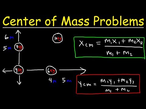
Center of Mass Physics Problems - Basic Introduction
 0:04:51
Mass Spectrometry
 0:04:37
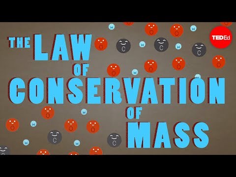
The law of conservation of mass - Todd Ramsey
 0:49:04
My problems with Vetra in Mass Effect Andromeda
 0:06:27
Mass Effect 3: Citadel - Technical Issues
 0:18:07
Moment, Center of Mass, and Centroid - Calculus Problems
 0:06:11
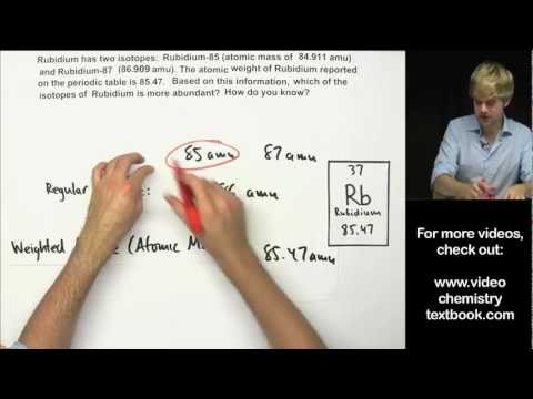
How to Calculate Atomic Mass Practice Problems
 0:04:07
Mass spec base peak example
 0:31:25
Molarity, Molality, Volume & Mass Percent, Mole Fraction & Density - Solution Concentration ...
 0:10:02
Mass Spectrometry
 0:11:53
Center of Mass
 0:13:07
Mass Spectrometry - Interpretation Made Easy!
 0:04:30
Calculate %m/m (Percent by Mass of a solution)
 0:07:46
Mass-Mass Stoichiometry
 0:13:11
How to Calculate Molar Mass Practice Problems
 0:08:07
Dog on a boat center of mass problem
 0:11:26
Stoichiometry Mass-Mass Problems
 0:00:55
Center Of Mass Concept |😀 #physicsexperiment 🙂#physics #scienceexperiment #arpitgupta #shorts #fun 👍...
 0:04:57
Centres of Mass : Toppling Problems : ExamSolutions
Комментарии