filmov
tv
#echo #heart #medical #diagnosis

Показать описание
An echocardiogram (often referred to as an echo) is a non-invasive medical test that uses ultrasound waves to create real-time images of the heart. It helps doctors assess the structure and function of the heart, including the heart valves, chambers, and blood flow.
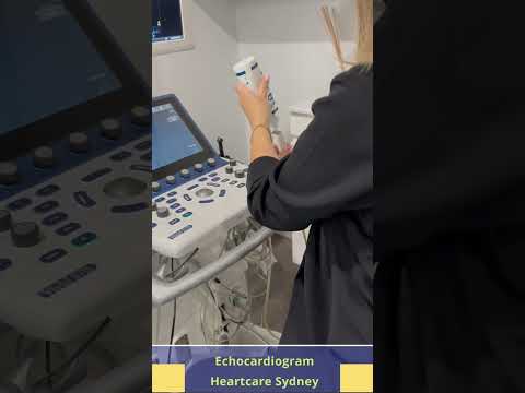
The procedure typically involves a gel being applied to the skin over the chest, followed by a small device called a transducer, which emits sound waves that bounce off the heart's structures and create images on a monitor.
An echocardiogram can be used to:
1. Evaluate heart function: Checking how well the heart pumps blood.
2. Assess heart valves: Identifying any problems with valve function or leakage.
3. Measure heart chamber size: Detecting abnormal enlargement or thickening.
4. Detect heart diseases: Such as heart attacks, heart failure, or congenital heart defects.
5. Monitor existing heart conditions: For ongoing assessment of heart disease management.
There are different types of echocardiograms, such as:
Transthoracic echocardiogram (TTE): The standard form where the transducer is placed on the chest.
Transesophageal echocardiogram (TEE): A more detailed echo where the transducer is inserted down the throat to get closer images of the heart.
Stress echocardiogram: Performed during or after exercise to evaluate the heart's function under stress.
It’s a safe and painless procedure, typically lasting between 30 minutes to an hour, and is commonly used to guide diagnosis and treatment for various heart-related conditions.
 0:00:06
 0:00:05
 0:00:05
 0:00:07
 0:00:06
 0:17:56
 0:00:05
 0:00:50
 0:00:33
 0:03:54
 0:00:57
 0:00:35
 0:00:59
 0:00:12
 0:00:05
 0:00:05
 0:00:16
 0:04:20
 0:00:17
 0:00:13
 0:01:20
 0:00:05
 0:03:18
 0:05:58